
Optics Mag is reader-supported. When you buy via links on our site, we may earn an affiliate commission at no cost to you. Read more.
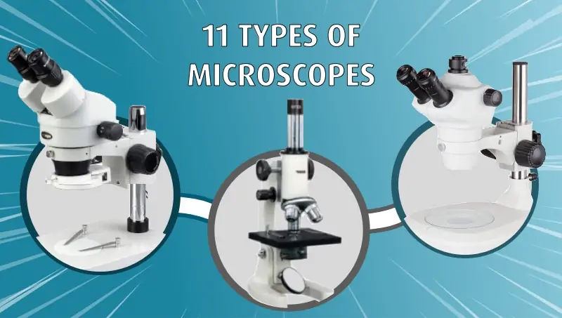
11 Different Types of Microscopes (With Pictures)
Last Updated on
Microscopy can change your perception of the world. We see everything in the limited spectrum of our eyes, and the micro world is more full of life than we often realize. Watching cells divide, seeing the structure of a hair follicle, or observing the intricate wings of an insect can both captivate and educate. But there is more than one way to view the microscopic world. You might switch on a compound microscope at home to observe tissue cells, or visit the world’s most powerful microscope at Lawrence Berkeley National Labs to see an image half the width of a hydrogen atom. We’ve broken down 9 of the most common microscope types below so you can learn more about these indispensable optical machines.

The 11 Types of Microscopes:
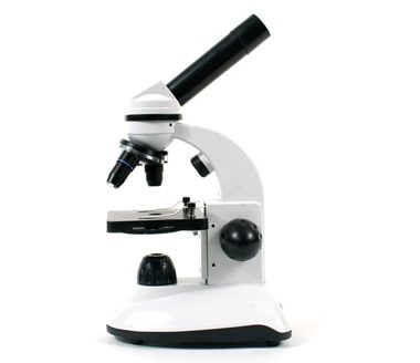
1. Light Microscopes
The most common type of microscope you’re likely to come across, these microscopes rely on lenses and light to illuminate a specimen for optimal image-gathering. They can be used for viewing living cells, insects, for performing dissections, or for clinical blood and tissue assessment.
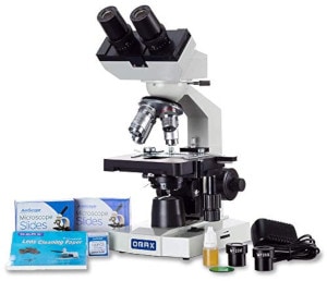
2. Compound Microscopes
You’ve undoubtedly peered into this type of microscope in your lifetime. Compound microscopes can be found in schools and labs across the world. They fit on a desktop, they’re portable, affordable, and easy to use. Their light source comes from the bottom, and light must pass through the specimen to travel through the microscope’s lenses and make it fully visible. They are most often used to view objects at a cellular level and can reach magnifications up to 1000x.
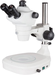
3. Stereoscopic Microscopes
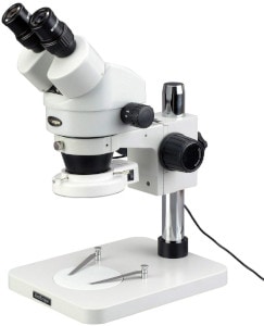
These are common in labs and educational settings, as well. A stereoscopic microscope has a light source on the top to illuminate the specimen, causing reflection into the microscope lens. They have weaker magnification than compound microscopes, to make it easier to see opaque, larger objects up close, at a maximum magnification of about 50x. Dual light paths inside the microscope tube create layered imaging, which provides a 3-dimensional image in the eyepiece, an improvement over the flat imaging in a compound scope. These are commonly used for dissection, coin appraisal, gem and mineral study, and entomology. They can also be used for intricate watch or microchip repair.
4. Confocal Microscopes
Confocal microscopes use lasers to scan a specimen and create high resolution, high magnification images. Because they provide depth-selection by scanning the specimen, they can create sectional detail (without physical dissection) that can be used to build a 3D image. Confocal microscopes are most often used in biomedical sciences to image living cells or embryos marked by fluorescence. They can typically reach a maximum magnification of 2000x.
5. Electron Microscopes
An electron microscope does not need light to create an image. Instead, this type of microscope sends accelerated electrons across or through a specimen to render a digital image. These microscopes have the highest power and highest resolution available and are used to see detailed structure at the cellular and macromolecular levels. While this may seem like the answer to all things microscopy, electron beams destroy samples. This means you can’t use them to view live specimens.
6. Scanning Electron Microscopes (SEM)
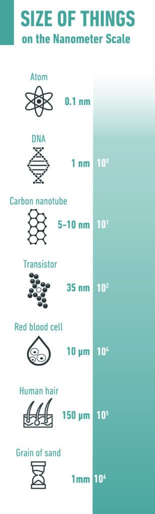
SEM microscopes scan the surface of a specimen in a rectangular pattern to provide information about topography and composition. The sample is set on a stage inside a vacuum chamber, which removes any electron-inhibiting air to aid acceleration. Information is then sent to a computer for interpretation and a digital image. SEMs can reach resolutions of about 10 nanometers, and have a maximum magnification strength of 30,000x.
7. Transmission Electron Microscopes (TEM)
Unlike the scanning structure of an SEM microscope, a TEM must pass electrons through a thin specimen to receive information, comparable to the way light must pass through a specimen on a compound microscope. Rather than reflecting off the specimen’s surface, the TEM’s electrons pass back and forth through the microscope’s vacuum chamber to build an image. Stronger than an SEM microscope, a TEM produces high magnification power of up to 1-nanometer resolution, or about 500,000x.
8. Reflection Electron Microscopes (REM)
These microscopes are used to study the microscopic surface structure and composition of crystals. A narrow beam of electrons is refracted from the first few atomic layers of the crystal at high resolution (up to about 1 nanometer). It’s combined with spectroscopy (the study of light dispersal) to form an image.
9. X-Ray Microscopes
Because X-rays can penetrate matter efficiently, they can be used to view the internal structure of opaque specimens such as rocks, bones, or metals. While lacking the power of an electron microscope, they don’t require a vacuum tube or accelerated electrons and so can handle any kind of specimen. X-ray microscopes can reach a resolution of about 20 nanometers.
10. Scanning Probes
SPMs can create nanoscale images at a resolution of less than 1 nanometer. A probe tip about as wide as a single atom scans across the specimen surface. It detects any deflections in the specimen and measures them via laser, then sends the information into photodiodes, which interpret the information into a digital image. These microscopes are used to study objects on a nanoscale and look inside cells and molecules.
11. Scanning Acoustic
These types of microscopes are used to image the internal structures of specimens without causing damage. They can achieve resolution down to 100 nanometers, are often used to inspect optical or electronic devices. Specimens are submerged in liquid and subjected to sound waves, which echo back to a transducer which pixelates the information and creates an image.

The Tiny World of Microscopy (and the Types of Microscopes to See it With)
Consider yourself lucky if you’ve had the opportunity to use each of these different microscopes. Some of them, such as electron microscopes, are so expensive that you will only find them in universities or laboratories. But the world of microscopy is growing every day, and the more technological advances are made in the field, the more the micro-world will be revealed.
We hope that this guide helps you find the right type of microscope for the needs you have.
More posts on microscopes:
About the Author Robert Sparks
Robert’s obsession with all things optical started early in life, when his optician father would bring home prototypes for Robert to play with. Nowadays, Robert is dedicated to helping others find the right optics for their needs. His hobbies include astronomy, astrophysics, and model building. Originally from Newark, NJ, he resides in Santa Fe, New Mexico, where the nighttime skies are filled with glittering stars.
Related Articles:
Monocular vs Telescope: Differences Explained (With Pictures)
How to Clean a Refractor Telescope: Step-by-Step Guide
How to Clean a Telescope Eyepiece: Step-by-Step Guide
How to Clean a Rifle Scope: 8 Expert Tips
What Is a Monocular Used For? 8 Common Functions
How to Clean a Telescope Mirror: 8 Expert Tips
Brightfield vs Phase Contrast Microscopy: The Differences Explained
SkyCamHD Drone Review: Pros, Cons, FAQ, & Verdict