
Optics Mag is reader-supported. When you buy via links on our site, we may earn an affiliate commission at no cost to you. Read more.
Brightfield vs Phase Contrast Microscopy: The Differences Explained
Last Updated on

While the concept of magnification is not new, its development and diversification are modern innovations. The Ancient Romans and Greeks observed its effects in nature. You can see them yourself by looking at something through a drop or glass of water, as the Roman philosopher Seneca observed about 2,000 years ago.1 The difference between brightfield and phase contrast microscopy is apparent once you take a closer look at the terms themselves.
The former describes the most common use of this technology and its resulting value. The latter’s main benefit is contrast. It allows you to see what the former does not, offering a detailed view that brightfield microscopy may not be able to provide. The difference lies in how the respective devices deal with the specimen’s reflective indices or contrast.2

Overview of Brightfield Microscopy:
Your first experience using a microscope was undoubtedly with a brightfield model. It represents the first generation of these devices, building upon centuries of development ongoing since the Dark Ages. Its invention was vital to later variations, such as phase contrast microscopy. It also paved the way for other uses and expanded its reach into other industries.
Surprisingly, we don’t know who invented the microscope, let alone the brightfield model. Many people were simultaneously working on its development in different countries and fields. Ironically, some did not have a formal science background, such as Roger Bacon and British amateur scientist Joseph Jackson Lister,3 who invented dark field microscopy.
How Brightfield Microscopy Works
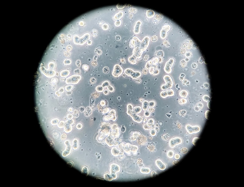
The concept of brightfield microscopy is simple yet brilliant. It builds on observations the Ancients made. A light source at the microscope’s base illuminates an object through a condenser on the bottom of the stage where the prepared slide sits.4 This setup lights it from the bottom. You can view the slide and its contents by looking through the ocular lens with magnification provided by the objective lens.
The light comes from a mirror you can adjust to catch the strongest angle. Other models use a powered source, whether it is from batteries or electricity. The difference affects the usability and visibility of the specimens you’re viewing.
Benefits of Brightfield Microscopy
Brightfield microscopy remains the most widespread use of this technology for a reason. It’s easy to use at an affordable price. It’s what you’ll find in products made for child use. It is an excellent summary of its benefits. You don’t need a sophisticated instrument to realize the value this microscope offers. That makes it a great choice for an entry-level model for school children to dip their toes into science and microbiology.

Disadvantages of Brightfield Microscopy
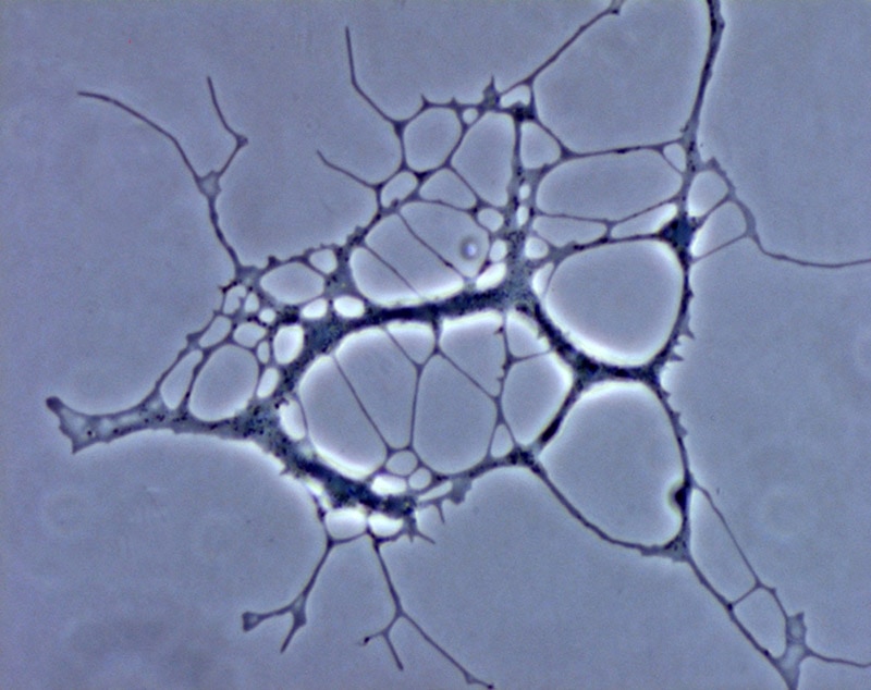
The microscope has limitations. It works best with low-contrast specimens because of this setup. That takes many biological samples off the table because some microorganisms are transparent, with little contrast between the structures within them. You’re left with using stains. These serve several purposes. They can fix a specimen to observe a specific life stage or development process. Unfortunately, they can also kill them.
Brightfield microscopy can either enhance or hinder what you’re trying to see. That’s why you must consider the type of technology you use with its supporting actors, like stains. These constraints spurred the development of other variations, including phase contrast microscopy.
Pros
- Easy to use
- Multiple options
- Affordable models available
- Useful for some live specimens
Cons
- Ineffective for low-contrast specimens
- Magnification limits

Overview of Phase Contrast Microscopy:
While it’s hard to overstate the value of brightfield microscopy, its drawbacks were evident in some applications, particularly biology. It was easy to view relatively larger specimens under magnification, such as insects. However, cells, Protista, bacteria, and other microscopic subjects were a different matter. It’s hard to differentiate microorganisms and structures without evident contrast. Everything looks the same.
Dutch physicist Frits Zernike began experimenting with ways to improve diffraction grating, which had just come online in the 1920s. This technology allows one to “see” non-visible things by separating light into its various wavelengths to provide a visible signature of different elements, not unlike a prism. Diffraction grating made it possible to view the atomic spectra of chemicals.
Zernike later connected his work and its use in microscopy using phase contrast. By chance, he learned that observing specimens in greater detail was possible with this technology. The German optics company produced the first phase contrast microscope in 1942. The Royal Swedish Academy of Sciences bestowed the 1952 Nobel Prize for Physics to Zernike for his remarkable achievement.
How Phase Contrast Microscopy Works
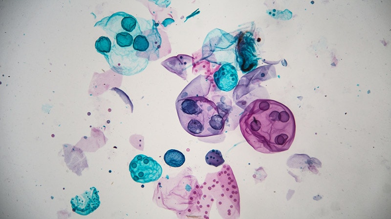
Phase contrast microscopy increases the reflective indices of a specimen by altering the light wavelengths. What looked like a solid-colored microorganism or cell now has depth and visible features. The minute differences in wavelengths allow the observer to view specimens that were impossible to see. Suddenly, you could look at living cells in action without using stains.
Phase contrast microscopy also allows for higher magnification of 400x and higher. To say that its uses were earth-shattering is a gross understatement. The science of biology took a massive step forward with its discovery.
Benefits of Phase Contrast Microscopy
Phase contrast microscopy broadened the application of this technology with benefits that extended far beyond the laboratory.
Pros
- Increased contrast
- Excellent visible detail
- Higher magnifications
- Revolutionary advancements in biology
Cons
- Expensive
- Typically professional grade only
Other Factors to Consider
An excellent starting point with any purchase is to begin with how you intend to use a device. The comparison between these two types of microscopy is a fitting example. We can boil it down to whether you simply want to magnify specimens or if seeing more structural detail is the goal. A decent brightfield microscope will probably suffice for most non-professional uses.
The Cost Factor
Magnification isn’t the only consideration when buying a microscope. You can get a handheld model for less than $50. You’re more likely to find an entry-level brightfield microscope for less than $100. When you start talking about a phase contrast device, you’re in four-figure or more territory. Most likely, if you’re buying a product for personal use, the latter is probably not an option, making the former a better choice.
Stereo vs Compound Microscope With Brightfield Technology
The next question you might have relates to what you’re most likely to view. If it’s relatively bigger items, like insects or plants, you’ll get your money’s worth with a stereo microscope. They typically have lower magnifications, usually up to 45x. A compound microscope will come in handy for observing small specimens, such as pond water. They are high-powered for these purposes.
Remember that magnification is a product of the spec that the eyepiece provides multiplied by the strengths of the objective lens. If the former is 10x and the latter 40x, the microscope allows you to see specimens 400 times bigger.
| When to Use a Brightfield Microscope | When to Use a Phase Contrast Microscope |
| Live specimens not harmed by stains | Live specimens not harmed by intense light |
| Using stains | Stains not optional |
| High contrast is not necessary | Details vital for use |
| Casual use | Medical applications |
Conclusion
Comparing brightfield and phase contrast microscopy provides a helpful introduction to the development of this technology. The latter opened up new worlds and possibilities no one could have imagined. Ironically, we benefit from both types every day without even knowing it. It also offers an excellent lesson in the power of small.
Featured Image Credit: (Left) Power J, Shutterstock (Right) Maple-Ferryman, Shutterstock
About the Author Chris Dinesen Rogers
Chris has been writing since 2009 on a variety of topics. Her motto with all of her writing is “science-based writing nurtured by education and critical thinking.” Chris specializes in science topics and has a special love for health and environmental topics, and animals of all shapes and sizes.
Related Articles:
What Is the Best Binocular Magnification for Hunting? Optical Features Explained
Monocular vs Telescope: Differences Explained (With Pictures)
How to Clean a Refractor Telescope: Step-by-Step Guide
How to Clean a Telescope Eyepiece: Step-by-Step Guide
How to Clean a Rifle Scope: 8 Expert Tips
What Is a Monocular Used For? 8 Common Functions
How to Clean a Telescope Mirror: 8 Expert Tips
SkyCamHD Drone Review: Pros, Cons, FAQ, & Verdict