
Optics Mag is reader-supported. When you buy via links on our site, we may earn an affiliate commission at no cost to you. Read more.
What is Brightfield Microscopy Used For? The Interesting Answer!
Last Updated on

Brightfield microscopy is a form of microscope illumination or a light microscopy technique. In brightfield microscopy, light is shined upwards from beneath the specimen, toward the microscope’s lens. The word “brightfield” in brightfield microscopy simply refers to the bright viewing field that shines against the darker specimen.
Brightfield microscopes are more commonly known as compound light microscopes.

How Does It Work?
Altogether, there are four light microscopy techniques. They are:
- Darkfield microscopy
- Phase microscopy
- Fluorescence microscopy
- Brightfield microscopy

Each of these techniques has its own advantages and uses. For example, phase microscopy is often used for viewing translucent images. In this article, we’re going to focus on how brightfield microscopy works.
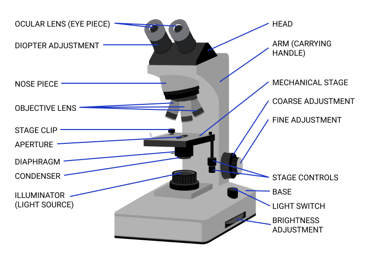
Brightfield microscopes have the following main components, in descending order:
- An ocular lens (or eyepiece)
- Objective lens (usually 3 or 4 that can be rotated into place)
- Mechanical stage
- Condenser lens, with diaphragm
- Illuminator (light source)
In brightfield microscopy, the specimen is placed on the mechanical stage. Directly beneath this stage is a lens, called a condenser lens, and beneath this is the microscope’s light source. When switched on, the light points up through the condenser lens and then the stage, towards the microscope’s objective lens.
Condenser Lens, Aperture, and Light
The condenser lens usually has an aperture diaphragm—a hole—that can be adjusted to control the contrast and resolution of the image. Generally, the diaphragm is opened more for thicker specimens or ones that have been dyed. However, opening the aperture diaphragm of the condenser lens will decrease the depth of field, which will require the microscope user to use the fine focus adjustment knobs to refocus.
If the aperture is entirely closed, the light will not be able to pass through, and the sample may appear too dark. That said, if it can be avoided, the condenser lens aperture should not be used to control the amount of light going through to the specimen—instead, the light dimmer should be used. For higher magnification, a very bright light source is needed, while low light is best for low magnification.
As light travels through and around the specimen, it’s collected through the microscope’s objective lens. The objective lens magnifies the light as it passes through, and eventually, the image reaches the eyepiece.
Where Is It Used?
Brightfield microscopy is used in many fields, from botany to microbiology. This technique can be used to observe and study bacteria, viruses, parasites, plant cells, blood cells, skin cells, and more.
Compound light microscopes are easily available, affordable, and simple to use. Because of their relative simplicity, brightfield microscopes are often one of the first microscopes students will use in schools and colleges.
Sometimes, specimens need to be stained for better contrast and viewing. For example, when examining thin slices of biological tissue, the specimen can be stained with Hematoxylin and Eosin. Hematoxylin turns the nuclei of the cells blue, while Eosin stains cytoplasm and other matter red or pink. This contrast of colors makes for easier examination.
With the addition of a polarizing filter, geologists can use brightfield microscopy to analyze rock and earth specimens closely.

Advantages of Brightfield Microscopy
Brightfield microscopy is, by far, one of the most accessible when it comes to cost and availability. Compound light microscopes can be obtained for all kinds of purposes, from educational toys to professional lab equipment, and everything in between.
These microscopes are also one of the easiest to use. They are versatile and can be used for many applications, ranging from studying pond water to bacteriology. Additional pieces of equipment (such as a polarizing filter) can be used to adapt brightfield microscopes for other uses too.
Disadvantages of Brightfield Microscopy
One of the disadvantages of brightfield microscopy is that it cannot be used to observe living bacteria, only fixed specimens. Another disadvantage is that the bright light source needed for good viewing can sometimes cause the specimen to become damaged through heat.
Brightfield microscopes have relatively low contrast, which means that many specimens need to be stained for proper viewing, a process that some people may find tedious.
Despite these disadvantages, brightfield microscopes are still commonly used throughout the scientific and educational sectors. Most hobbyists begin with compound light microscopes, too.
Frequently Asked Questions (FAQs)
What is brightfield microscopy used for?
Brightfield microscopy is used to examine a wide variety of specimens and samples across many disciplines, including microbiology. Amongst many other uses, they can be used to identify bacteria and examine tissue samples. Geologists can use them to find otherwise invisible characteristics in rock and earth samples. Botanists use brightfield microscopes to study plants. These microscopes can also be used to examine pond water.
What are the characteristics of a brightfield microscope?
Brightfield microscopes have an incandescent light source at the base, pointing up. They usually have a “wheel” consisting of 2 to 5 objective lenses with differing magnifications, that can be rotated into place. Beneath the sample stage, there is usually a diaphragm that can be adjusted to focus the light.
What is the difference between brightfield and darkfield microscopy?
Whereas in brightfield microscopy, the specimen appears dark while the background is bright, in darkfield microscopy, the opposite is true—the specimen is bright against a dark background.
The advantage of darkfield microscopy is that staining is usually not required. Whereas in brightfield microscopy, only fixed specimens can be viewed, in darkfield microscopy, both living and fixed specimens can be examined.
The disadvantage of using darkfield microscopy over brightfield is that only the external structure of the specimen will be visible, and the internal structure will not. Compared to brightfield microscopy, darkfield microscopy usually costs more.
What is the difference between brightfield and phase contrast microscopy?
The optics of phase contrast microscopes transform the differences in the refractive index of materials into differences in brightness. Even translucent specimens will appear brighter or darker than the background without the need for staining, unlike in brightfield microscopy—where the specimen would need to be killed, fixed, and stained before viewing.

What is the advantage of phase contrast microscopy over brightfield microscopy?
Phase contrast microscopy can be expensive, but the advantage of using this technique of microscopy over brightfield microscopy is that live specimens can be viewed, and at very high magnification. Unlike darkfield microscopy, both the internal and external structure of the specimen can be observed in great detail during phase contrast microscopy.
Can plants be studied with brightfield microscopy?
Plant specimens can be viewed directly with brightfield microscopy, without the need for staining. Cross sections of fruits, leaves, petals, stems, and vegetation can all be viewed using a brightfield microscope. It can also be used to successfully study thin slices of tree branches, bark, wood, roots, and even cross-sections of spruce and pine needles.
Summing Up
Brightfield microscopy is one of the most common light microscopy techniques in the world. It involves shining a bright light through a specimen towards the microscope’s objective lens. The technique allows the specimen to absorb some light, so that it appears darker against the “bright field” in the background.
Brightfield microscopy has many applications, ranging from microbiology to botany and geology. Commonly known as compound light microscopes, these microscopes are easy to use and widely available, making them a perfect choice for beginners and pros alike.
See also:
- What Is a Parfocal Microscope? Pros, Cons and How It Works
- What Is a Scanning Electron Microscope (SEM)? How Does It Work?
- Brightfield vs Phase Contrast Microscopy: The Differences Explained
Featured Image Credit: AndriyShevchuk, Shutterstock
Table of Contents
- How Does It Work?
- Where Is It Used?
- Advantages of Brightfield Microscopy
- Disadvantages of Brightfield Microscopy
- Frequently Asked Questions (FAQs)
- What is brightfield microscopy used for?
- What are the characteristics of a brightfield microscope?
- What is the difference between brightfield and darkfield microscopy?
- What is the difference between brightfield and phase contrast microscopy?
- What is the advantage of phase contrast microscopy over brightfield microscopy?
- Can plants be studied with brightfield microscopy?
- Summing Up
About the Author Cheryl Regan
Cheryl is a freelance content and copywriter from the United Kingdom. Her interests include hiking and amateur astronomy but focuses her writing on gardening and photography. If she isn't writing she can be found curled up with a coffee and her pet cat.
Related Articles:
What Is the Best Binocular Magnification for Hunting? Optical Features Explained
How to Clean a Refractor Telescope: Step-by-Step Guide
How to Clean a Telescope Eyepiece: Step-by-Step Guide
How to Clean a Rifle Scope: 8 Expert Tips
Monocular vs Telescope: Differences Explained (With Pictures)
What Is a Monocular Used For? 8 Common Functions
How to Clean a Telescope Mirror: 8 Expert Tips
Brightfield vs Phase Contrast Microscopy: The Differences Explained