
Optics Mag is reader-supported. When you buy via links on our site, we may earn an affiliate commission at no cost to you. Read more.
What Is a Scanning Electron Microscope (SEM)? Pros, Cons & FAQs
Last Updated on

Scanning electron microscopes use electrons instead of light to form an image. They’ve been in development since the early 1950s, offering new possibilities in the realm of medicine and physical sciences. It was designed to directly study the surfaces of solid objects using a beam of focused electrons of low energy.
These microscopes may be used across a number of industrial, commercial, and research applications, including forensics and high-level fabrication.

How Does It Work?
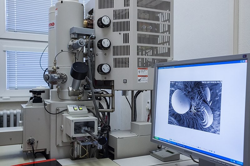
The SEM is a microscope that produces a highly magnified image with electrons instead of light. An electron beam is produced at the top of the microscope with an electron gun, which travels through the microscope in a vacuum tube. The beam travels through electromagnetic fields and lenses to focus it on the sample. Once it hits the sample, the electrons are ejected from the sample. Detectors collect the electrons and convert them into a signal that’s then carried to a screen, producing the final image.

What Are the Differences Between Scanning Electron Microscopes and Light Microscopes?
With sufficient light, the human eye can distinguish two points in space up to 0.2 mm apart without additional lenses. This is the resolution of the eye. Microscopes use an assembly of lenses to magnify the distance and allow the human eye to see points much closer together.
A light microscope has a maximum magnification of about 1000x. This is limited by the number and quality of the lenses, as well as the wavelength of light used for illumination. Electron microscopes have shorter wavelengths, allowing better resolution.
The dimensions for materials and devices are shrinking, reaching the limit of what light microscopy can accomplish. Electron microscopy is required to see many of these materials and devices, such as a nanofiber layer or asbestos samples.
Where Is It Used?
Scanning electron microscopes offer a world of possibilities for industrial, commercial, and research use. In materials science, scanning electron microscopes may be used for research, quality control, and failure analysis for superconductors, nanotubes, nanofibers, and more. They’re also integral to microchip assembly and production, which is becoming increasingly important as IoT and other emerging technologies see increased adoption.
In forensics, scanning electron microscopes are essential for gathering forensic insights like ballistics comparisons, examination of currency authenticity, paint and fiber analysis, gunshot residue analysis, and filament bulb analysis for traffic accidents. In the biological sciences, these microscopes allow researchers to test vaccines, identify new pathogens, discover new species, and perform work in the field of genetics.
The scanning electron microscope has many advantages over light microscopy, including no elaborate specimen preparation and the ability to accommodate large and bulky specimens. This technology may be combined with transmission electron microscopes, forming a scanning transmission electron microscope that may be used to study thick sections with higher degrees of contrast and brightness.
Advantages of Scanning Electron Microscopes
Few instruments compare to the capabilities of scanning electron microscopes in studying solid materials. They’re used in medicine, biology, physical sciences, forensics, materials sciences, and more, offering greater resolution than other instrumentation. These microscopes are also easy to operate and feature intuitive interfaces, and the data acquisition is quick. Most modern scanning electron microscopes provide data in digital formats, which are easy to transport to different programs or remote teams.
Disadvantages of Scanning Electron Microscopes
Scanning electron microscopes do have some disadvantages, however. The material samples must be solid and small enough to fit into the microscope chamber. The materials must be stable in the vacuum, and any samples likely to outgas at low pressures are unsuitable for use with scanning electron microscopes. These may include wet samples like swelling clays or coal.
In addition, the EDS detectors on scanning electron microscopes aren’t able to detect light elements like lithium, hydrogen, and helium, nor can they detect elements with atomic numbers lower than 11.
Frequently Asked Questions (FAQs)
Should I use optical microscopy or scanning electron microscopy?
Optical microscopy and scanning electron microscopy have appropriate uses and pros and cons. Scanning electron microscopes are superior in resolution and depth of focus, but optical microscopes are easier and faster to use for quick, coarse study. For example, a light microscope may be used to detect major defects, then the scanning electron microscope can be used to observe defects in greater detail.
How are microscopy samples prepared?
Scanning electron microscopes use vacuum chambers and electrons to form an image, so the samples must be prepared specially. All water has to be removed since it would vaporize in a vacuum. Metals are conductive and require no preparation, but non-metals need to be prepared with a thin layer of conductive material.
To prepare the sample, preparation instruments like critical point dryers and freeze dryers are designed to remove water from biological and liquid-based substances. Most biological specimens require chemical stabilization to preserve them and allow them to withstand subsequent drying and coating processes.
What is cryo preparation?
Cryo preparation techniques for scanning electron microscopes are now considered essential for wet specimens. This removes the need for other preparation methods, including chemical fixation and critical point drying to allow the specimen to be observed in its natural hydrated state. It also reduces the other issues with conventional preparation, including shrinkage and distortion, mechanical damage, and toxic reactions.
Are scanning electron microscopes safe?
Yes, scanning electron microscopes are safe with proper training and safety procedures. The safety concerns are related to the electrons that are backscattered from the sample, as well as the X-rays produced in the microscopy process. Most scanning electron microscopes are well shielded and do not produce significant exposure rates, but they need to be monitored for shield integrity.
A Quick Reference Guide
| Scanning Electron Microscopy | Light Microscopy |
| Gray-scaled image | Natural color |
| Higher resolution | Lower resolution |
| Better depth of focus | Lower depth of focus |
| High magnification capability | Low magnification for quick, coarse study |
Light microscopy and electron microscopy have advantages and disadvantages for different applications. For many applications, light microscopes are more accessible and easier to use, making them ideal unless the research calls for better resolution, more depth of focus, or higher magnification.
Summing Up
Scanning electron microscopes offer virtually endless applications for industrial and research uses. With the increasing focus on quality control at microscopic scales, having high-resolution imagery with a scanning electron microscope provides deeper insights. When combined with transmission electron microscopy, scanning electron microscopes are arguably the most powerful and capable instruments we have for examining solid-state materials.
You Might Also Be Interested In:
- What Is a Parfocal Microscope? Pros, Cons and How It Works
- What is Brightfield Microscopy Used For? The Interesting Answer!
Featured Image Credit: Elizaveta Galitckaia, Shutterstock
About the Author Robert Sparks
Robert’s obsession with all things optical started early in life, when his optician father would bring home prototypes for Robert to play with. Nowadays, Robert is dedicated to helping others find the right optics for their needs. His hobbies include astronomy, astrophysics, and model building. Originally from Newark, NJ, he resides in Santa Fe, New Mexico, where the nighttime skies are filled with glittering stars.
Related Articles:
Binocular Magnification Chart: Numbers & Distances Compared
What Is the Best Binocular Magnification for Hunting? Optical Features Explained
When Were Binoculars Invented? History, Today & Future
How to Clean a Refractor Telescope: Step-by-Step Guide
How to Clean a Telescope Eyepiece: Step-by-Step Guide
How to Clean a Rifle Scope: 8 Expert Tips
Monocular vs Telescope: Differences Explained (With Pictures)
What Is a Monocular Used For? 8 Common Functions