
Optics Mag is reader-supported. When you buy via links on our site, we may earn an affiliate commission at no cost to you. Read more.
Transmission (TEM) vs Scanning (SEM) Electron Microscopes: What’s the Difference?
Last Updated on

Electron microscopes allow the viewer to see objects that are so small that no amount of light-based magnification can make them visible. Light particles (photons) travel in waves, and like anything else that travels in waves, photons have a certain wavelength. If an object is physically smaller than the wavelength of a photon, that object will never be visible if you are using lenses, prisms, or anything else to focus the photons.
Enter the electron microscope. The basic idea behind an electron microscope is to use electrons to hit or pass through the object instead of photons, then have the machine inside convert the information into a visible image as if it had been made with photons. The best first step to understanding the differences between a TEM and an SEM is to understand how regular microscopes differ.

Light-Based Microscopes | Compound and Dissecting
There are two main ways that light is used in regular microscopes to magnify an image. Light can be used to pass through an image and up into the lenses, or light can be bounced off an image and back up into the lens of the microscope.
The two different methods come with some trade-offs. Passing light through an image allows you to achieve greater magnification and see more detail, but also requires your specimen to be at least somewhat translucent, and may involve preparation of the specimen with dyes or stains.
Bouncing light off an image limits your magnification but gives you a large enough working area to manipulate the specimen and interact with it while viewing it. It also results in a 3D image without the need to stain or discolor the specimen. Now, let’s talk about electron microscopes.
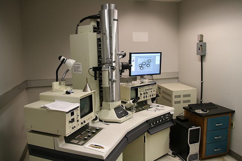
Overview of Transmission Electron Microscopes
A TEM could be considered the electron version of a compound microscope, which requires light to pass through the specimen and up into the lens. A TEM receives a beam of electrons after they pass through the object. The quantity and density of the electrons that make it through the specimen without getting stopped is translated into lighter and darker parts of the resulting image.
For this reason, specimens need to be thin enough to allow electrons to pass through them. How thin this is actually depends on how much energy the electron has, but it is generally less than 300 nanometers. Nanometers are incredibly tiny. To put it into perspective, a nanometer is .00000001 Centimeters, or in other words, 1 centimeter is equivalent to 10,000,000 nanometers.
How a TEM Works

After the specimen is prepared on a film that is thin enough (no easy task), it is placed in the microscope between the electron emitter and the electromagnetic lenses. With such small imaging, even the atoms between the specimen and the lens will obfuscate the image, so the space between the specimen and the lens must be a vacuum, which allows the electrons to travel freely without disruption.
The electrons hit the lens, and then the computer inside translates the information about how many electrons are hitting each part of the lens into visual information that allows intricate details to be seen on items as small as viruses, atoms, etc.
What a TEM Is Used For

A TEM is used to see the smallest of the small. Through a TEM, details as tiny as a single nanometer can be seen and analyzed. TEMs are most often used to image and analyze the structure of cells and molecules, so can be used to compare how those structures change over time or when affected by something else.
A TEM is used, much like a compound microscope, for when maximum magnification and seeing the details inside a specimen is most critical. The images that come off a TEM are black-and-white and two-dimensional, which makes them most useful for scientific research.
Pros
- Magnification down to a single nanometer
- Able to see inside specimens
- Higher resolution images
Cons
- More difficult and demanding to use
- Requires incredibly thin specimens
- 2-dimensional, black-and-white image
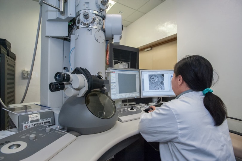
Overview of Scanning Electron Microscopes

An SEM, on the other hand, is much more similar to a dissecting microscope both in function and in purpose. An SEM bounces a stream of electrons off of the specimen and back up into the electromagnetic lens. This works best with specimens that are thick enough not to let all of the electrons pass through them.
Since you are not passing electrons through the specimen, it doesn’t have to be nearly as thin. This allows you to view different types of specimens and view different characteristics of them. An SEM gives you a 3D image but does not allow you to see inside of a specimen the way a TEM does. The 3D image from an SEM is more interesting than that of a TEM, but it can only view details down to around 15 nanometers.
Fifteen nanometers may not seem much bigger than 1 nanometer, but at the atomic and molecular level, that difference is significant.
How an SEM Works

A scanning electron microscope has the electron emitter on the same end of the vacuum tube as the electromagnetic lens and bounces the electrons off the surface of the specimen, which then bounce back up the tube into the lens. On the side is a secondary electron detector, which provides a lot of additional information about the surface of the specimen.
The image is then constructed using the data from the backscattered electron detector (the primary detector placed directly above the sample) and the secondary electron detector and shown on a digital screen in its magnified form.
What an SEM Is Used For

Most of the use cases for an SEM involve analyzing material compositions and structures, like fabric, silicon, or even sheet metal. SEMs can be used in developing new composites, analyzing existing ones, and measuring the effects of an experiment on a material. Often, this involves analyzing the cellular structure of a material, and SEMs are often used to better document and understand crystalline structures.
SEMs are found more often in engineering, industrial, and commercial applications than TEMs, since the information that engineers and materials scientists need usually does not require the increased magnification, nor do the materials they are working with lend themselves well to the functional limitations of a TEM.
Pros
- 3D image
- Easier to operate and interpret results
- More applicable outside of academia
Cons
- Cannot see inside specimens
- Does not magnify as much as TEM
The Cost Factor
Both TEMs and SEMs are prohibitively expensive, but a TEM is even more expensive, and far more training and expertise are required to successfully operate a TEM than an SEM. The cost of the machine itself is just a portion of the cost of operating an electron microscope. Technicians that are trained on the proper use of the microscope, electron emitters that need to be replaced much like light bulbs, and the electricity to run the machines all add to the cost.
Convenience and Ease of Use
Transmission electron microscopes won’t fill a room, but they are wider and taller than a scanning electron microscope. Neither are small, and the higher quality you buy the larger they will likely be. In most laboratories, the electron microscope will have a dedicated desk or countertop with space for a computer next to it.
Once a specimen is prepped, a TEM is easy enough to use for a trained technician. Prepping the specimen, however, can be far more complicated. For use in an SEM, the specimen still must be prepped but it is a far simpler process and can be done without as much training. An SEM is just as easy to operate once the specimen is prepped as a TEM.
When To Use Which?
If you need to see an item with the absolute maximum amount of magnification possible, then a transmission electron microscope is the right choice. If the type of specimen you are looking at is (or can be sliced to be) thin enough to allow electrons to pass through it, then a TEM will give you a much closer look than an SEM.
If you need to understand details on the surface of a material or other specimen, then an SEM will be the right choice. If you do not need to see inside cells or are working with a specimen that will not allow electrons to pass through it, then an SEM will do a much better job for you than a TEM.
Conclusion
Electron microscopes are not hobby devices; they are used only for extreme magnification of objects so tiny that visible light cannot capture them. A transmission electron microscope places the specimen between the electron emitter and the lens so that electrons must pass through the specimen to be visible in the resulting image.
A scanning electron microscope bounces electrons off the surface of the image and back into the lenses, which allows for a 3D image but does not resolve quite as much detail. Both of these devices are incredible pieces of ingenuity and have contributed immensely to science since they were invented.
About the Author Robert Sparks
Robert’s obsession with all things optical started early in life, when his optician father would bring home prototypes for Robert to play with. Nowadays, Robert is dedicated to helping others find the right optics for their needs. His hobbies include astronomy, astrophysics, and model building. Originally from Newark, NJ, he resides in Santa Fe, New Mexico, where the nighttime skies are filled with glittering stars.
Related Articles:
How to Clean a Refractor Telescope: Step-by-Step Guide
How to Clean a Telescope Eyepiece: Step-by-Step Guide
How to Clean a Rifle Scope: 8 Expert Tips
Monocular vs Telescope: Differences Explained (With Pictures)
What Is a Monocular Used For? 8 Common Functions
How to Clean a Telescope Mirror: 8 Expert Tips
Brightfield vs Phase Contrast Microscopy: The Differences Explained
SkyCamHD Drone Review: Pros, Cons, FAQ, & Verdict