
Optics Mag is reader-supported. When you buy via links on our site, we may earn an affiliate commission at no cost to you. Read more.
What Is a Transmission Electron Microscope? How Does It Work?
Last Updated on

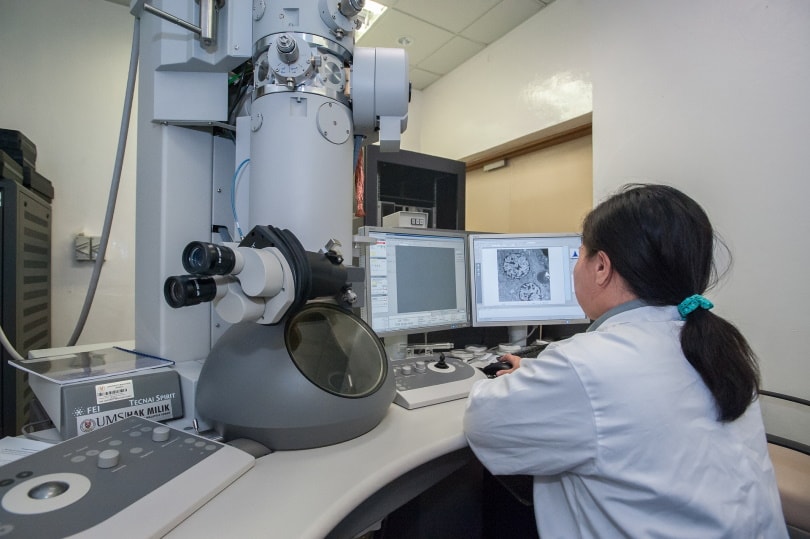
A transmission electron microscope is a type of microscope that uses electron particle beams to generate a highly magnified image for viewing specimens.1 It can magnify objects up to 2 million times, allowing images of ultrathin specimens like tissue sections and molecules. The image is then magnified and focused onto an imaging device, such as a layer of photographic film or a fluorescent screen.
Transmission electron microscopes are vital to the medical and biological fields to visualize objects like protein molecule structures, cell interiors, cytoskeletal filaments, and molecules in viruses.2 Though they have limitations, they’re a necessary component of ongoing research.

How Does It Work?
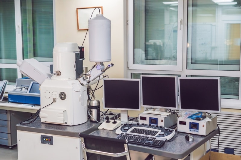
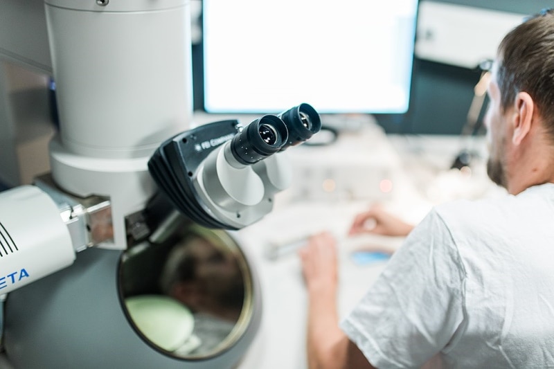
Transmission electron microscopes use a tungsten filament to create a high-voltage electron beam that travels through a vacuum chamber. The emitted electrons are accelerated through the electromagnetic field that focuses them into a fine beam, which is passed through the sample material. The sample is prepared to be thin—less than 100 nm—allowing the electrons to pass through.
As the electrons pass through the sample, they either scatter or reach a phosphor screen or charge-coupled device, or a camera that takes still or moving pictures of the specimen. The electrons may also terminate at a film that produces an image.
When the sample has less density, more electrons can pass through to produce a brighter image. Darker images are produced when samples are denser, allowing fewer electrons to get through. This is why it’s so important for the samples to be ultrathin during preparation. The image is sorted into different shades based on its density.
Differences Between Transmission Electron Microscopes and Light Microscopes:
Transmission electron microscopes and light microscopes have the same basic principles of operation, but beyond that, they’re quite different. As the name implies, transmission electron microscopes rely on electrons instead of light for magnifying images.
Light microscopes are limited by the wavelength of light and can only magnify up to 2,000 times. Electron microscopes, such as transmission electron microscopes, produce images with higher magnification because the beam of electrons have smaller wavelengths, leading to images with higher resolution.
Where Is It Used?
As mentioned, transmission electron microscopes are vital to the medical and biological fields. The high degree of magnification allows researchers to learn more about the structure, morphology, stress, and crystallization of different specimens.
This technology can be leveraged in many fields and industries, including materials science, gemology, forensic science, and epidemiology. Transmission electron microscopy is an essential component of virology and can enhance diagnostics and provide rapid treatment.
Advantages of Transmission Electron Microscopes
Transmission electron microscopes offer many of the same advantages as electron microscopy. The techniques offer enhanced material analysis with the capability of producing high-quality images. Transmission electron microscopy offers the highest magnification in the microscope field, offering numerous applications in scientific and industrial fields. With the high degree of magnification, transmission electron microscopes can yield vital information about a sample’s shape, size, structure, and surface features.
When used in combination with energy dispersive X-ray spectroscopy and electron energy loss spectroscopy, transmission electron microscopes can provide elemental and atomic-binding information for more comprehensive data.
Disadvantages of Transmission Electron Microscopes
There are disadvantages and limitations to transmission electron microscopes. The instruments are large and expensive, limiting access to them for some researchers. They must be housed and maintained in a specific manner to protect them from the fluctuation of electromagnetic fields, mechanical vibration, and the variation of cooling water. Special training is needed to prepare samples and operate these microscopes.
In addition, preparing the samples can be challenging and cumbersome, especially in bulk. It’s easy for sample preparation to be compromised by artifacts, and the specimens are limited to materials that can be prepared in a small enough sample to fit in the chamber. The materials must also tolerate the environment of a vacuum chamber.

Frequently Asked Questions (FAQs)
Why use a transmission electron microscope?
Questions involving samples that require high-resolution imagery, such as the structure of bacteria and viruses, the morphology of cells, and the characteristics of diseased tissue, can be answered with a transmission electron microscope. Other forms of microscopy do not offer the same degree of resolution.
What are the challenges of using transmission electron microscopes?
Transmission electron microscopes require precise environments and operations to yield valid results. These microscopes are sensitive to changes in their environments like vibration and fluctuating electromagnetic fields, so they must be set up and operated in strictly monitored and controlled areas. They also require a lot of maintenance to operate effectively.
How are specimens prepared for transmission electron microscopy?
Specimens must be ultrathin for transmission electron microscopy to allow electrons to pass through the material. Samples are prepared by slicing thin sections of a specimen’s tissue with a special tool called an ultramicrotome.
Samples with tissue must be submerged in a chemical solution to preserve cell structure and then must be dehydrated. Samples are then placed in hard, clean plastic for support during slicing. After slicing, sections of the sample are mounted on grids and stained with a solution of lead. This offers contrast to highlight certain cell parts. Once the sample is viewed with a transmission electron microscope, the electrons are scattered by the lead and do not penetrate the tissue, leaving them dark.
Errors during preparation can compromise the integrity of the sample and by extension, the results. This is why it’s so important for personnel to have the specialized training necessary to prepare transmission electron microscopy samples.
A Quick Reference Guide: Transmission Electron Microscopy vs Light Microscopy
| Transmission Electron Microscopy | Light Microscopy |
| Uses electron beams for imaging | Uses light for imaging |
| Higher magnification | Lower magnification |
| Specialized specimen preparation | Simpler specimen preparation |
| Only deceased and dry specimens may be used | Live and deceased specimens may be used |
| Image received on a fluorescent screen | Image is seen through an ocular lens with no screen necessary |
| Image formation depends on light absorption | Image formation depends on electron scattering |
| High resolution | Low resolution |
| Expensive and difficult to maintain | Affordable and low maintenance |
| Direct magnification up to 1,000,000x | Magnification of 500x to 1,500x |
Both transmission electron microscopes and light microscopes have their value in research, but electron microscopes are increasingly used to study specimens with high resolution. Light microscopes are still useful for educational environments and for viewing specimens that must be alive, and they’re widely available and easier to maintain than transmission electron microscopes.

Final Thoughts
Among the different types of microscopes, transmission electron microscopes have widened our research and data in fields like epidemiology, biology, and forensic science. As science asks deeper and more challenging questions, transmission electron microscopes and their high-resolution images are necessary to find the answers and fuel more research and scientific curiosity.
Featured Image Credit: Elizaveta Galitckaia, Shutterstock
Table of Contents
- How Does It Work?
- Differences Between Transmission Electron Microscopes and Light Microscopes:
- Where Is It Used?
- Advantages of Transmission Electron Microscopes
- Disadvantages of Transmission Electron Microscopes
- Frequently Asked Questions (FAQs)
- A Quick Reference Guide: Transmission Electron Microscopy vs Light Microscopy
- Final Thoughts
About the Author Robert Sparks
Robert’s obsession with all things optical started early in life, when his optician father would bring home prototypes for Robert to play with. Nowadays, Robert is dedicated to helping others find the right optics for their needs. His hobbies include astronomy, astrophysics, and model building. Originally from Newark, NJ, he resides in Santa Fe, New Mexico, where the nighttime skies are filled with glittering stars.
Related Articles:
What Is the Best Binocular Magnification for Hunting? Optical Features Explained
How to Clean a Refractor Telescope: Step-by-Step Guide
How to Clean a Telescope Eyepiece: Step-by-Step Guide
How to Clean a Rifle Scope: 8 Expert Tips
Monocular vs Telescope: Differences Explained (With Pictures)
What Is a Monocular Used For? 8 Common Functions
How to Clean a Telescope Mirror: 8 Expert Tips
Brightfield vs Phase Contrast Microscopy: The Differences Explained