
Optics Mag is reader-supported. When you buy via links on our site, we may earn an affiliate commission at no cost to you. Read more.
What Does DNA Look Like Under a Microscope? (With Pictures!)
Last Updated on

Most people are familiar with deoxyribonucleic acid (DNA) from illustrations and animations—a swirly strand of colorful fibers in reds, blues, and yellows. This rainbow, we are told, holds key genetic information and instructions for protein synthesis.
DNA under a microscope isn’t nearly as artistic. In fact, we can’t get the detail we imagine with DNA under a microscope, no matter how powerful, but we are making great strides in the right direction.

Can You See DNA Under a Microscope? What Does It Look Like?
Scientists may use electron, scanning tunneling, and atomic force microscopes to view individual DNA molecules, but these methods don’t provide an abundance of detail.
What does it look like? A string, basically. It is difficult to see DNA as the individual units that comprise it, for the most part. If the right stain is used, the spiral strands of chromosomes can be viewed through a powerful microscope.
But chromosomes contain tens of thousands of genes and infinitesimal details, which can’t be seen through microscopy. In other words, you can’t tell the difference in the DNA of two people with a microscope.
When Watson and Crick identified DNA’s structure, it was based on a bit of guesswork and some X-ray crystallography. The rendering was inexact but served to give us a visual for DNA strands.
The key is understanding how crystallography works. The imaging relies on diffracted light, so when we see an image of the DNA’s signature double helix, we’re not really looking at the DNA—we see the X-rays deflected from atoms.
The actual image of DNA under a microscope may not be as colorful as our DNA illustrations, but it’s realistic.
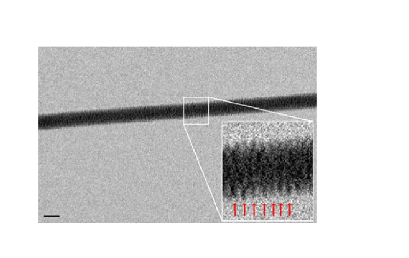
The image of a single thread of double-stranded DNA was created by Enzo di Fabrizio and his team at Italy’s University of Genoa, which developed an entirely new technique to capture an image of DNA. The team suspended DNA threads out of a dilute solution and laid them on a bed of nanoscopic silicon pillars, like tying thread on a bed of nails.
The pattern of the pillars was water-repellant, allowing the water to evaporate rapidly and leave the strands of DNA stretched, suspended, and ready to image. Tiny holes were also drilled into the base of the nanopillar bed, allowing beams of electrons to pass through and create resolution images of the corkscrew thread of the DNA double helix.
Using an electron microscope, the team managed to get a high-resolution image of a DNA thread to visualize what we’ve only seen in animations or slightly inaccurate crystallography images previously.

What Does DNA Look Like to You?
DNA under a microscope doesn’t offer the detail and resolution we imagine, like a cell structure, textile fibers, or highly magnified dust. We can see in Fabrizio’s electron microscopy image that a single strand contains all the wonders of genes, which may look like a strand of thread, ultrathin noodles, or delicate fibers. As microscopy advances, however, we may be able to see the individual double helices or an unwound single strand of DNA eventually.
Featured Image Credit By: Edgloris Marys, Shutterstock
About the Author Robert Sparks
Robert’s obsession with all things optical started early in life, when his optician father would bring home prototypes for Robert to play with. Nowadays, Robert is dedicated to helping others find the right optics for their needs. His hobbies include astronomy, astrophysics, and model building. Originally from Newark, NJ, he resides in Santa Fe, New Mexico, where the nighttime skies are filled with glittering stars.
Related Articles:
How to Clean a Refractor Telescope: Step-by-Step Guide
How to Clean a Telescope Eyepiece: Step-by-Step Guide
How to Clean a Rifle Scope: 8 Expert Tips
Monocular vs Telescope: Differences Explained (With Pictures)
What Is a Monocular Used For? 8 Common Functions
How to Clean a Telescope Mirror: 8 Expert Tips
Brightfield vs Phase Contrast Microscopy: The Differences Explained
SkyCamHD Drone Review: Pros, Cons, FAQ, & Verdict