
Optics Mag is reader-supported. When you buy via links on our site, we may earn an affiliate commission at no cost to you. Read more.
What Is an Electron Microscope? What Is Electron Microscopy?
Last Updated on

Scientific equipment has evolved quite rapidly over the past century, and electron microscopes are surely part of this sentiment. But have you ever wondered what an electron microscope is? Have you ever wondered what they’re used for? Electron microscopy, of course!
But what is electron microscopy anyway? In short, electron microscopes use electron particles instead of light to create detailed images. To answer those questions, we’ve created a handy guide to explain what electron microscopes are and how they work!

How Does It Work?
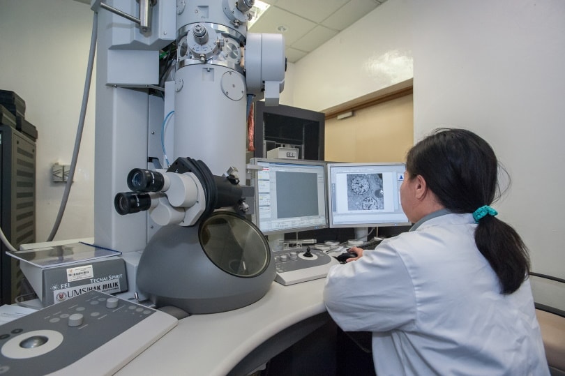
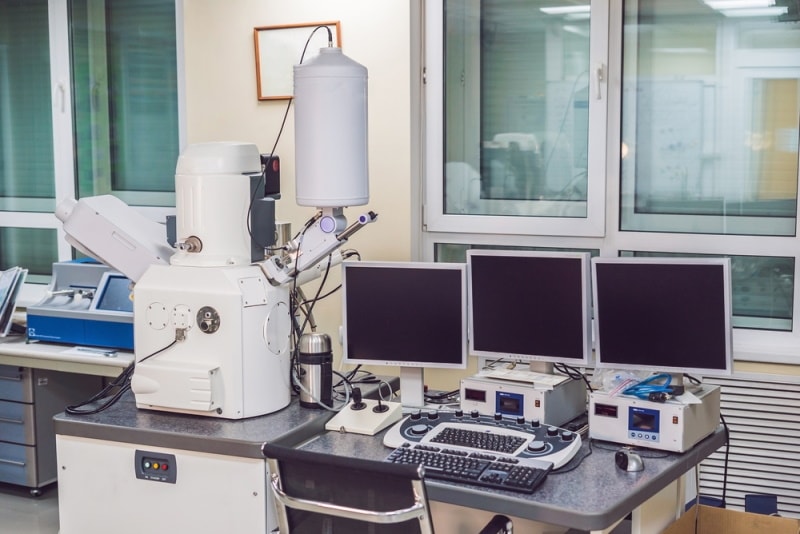
Electron microscopes are a type of microscope that takes advantage of electron particles rather than artificial lighting. With an ordinary microscope or compound microscope, light is the key player in resolving detailed images of a sample. In contrast, electron microscopes use electrons to scan subjects to create high-resolution images and are far more powerful. This is because electrons have a shorter wavelength compared to photons. They are also much less common due to their incredibly high cost and massive physical construction.
In 1931, physicist Ernst Ruska and engineer Max Knoll built the first-ever electron microscope, but it was just a prototype. However, their design led to a deeper understanding of this technology and how it could be used to advance microbiology studies. In fact, it didn’t take long for it to overtake the strength of a light microscope.
What Are the Different Types of Electron Microscopes?
With electron microscopes, there are generally two different kinds; scanning EM (SEM) and transmission EM (TEM). The “EM” in both SEM and TEM stands for electron microscopy.
SEMs are similar to your typical compound light microscope, but they are specialized for observing processes such as proteins or cells. They work by moving electrons back and forth across the surface of the sample, which creates an image that can be observed in great detail. SEMs are the primary tool that enables us to study bacteria or viruses such as Salmonella.
TEMs, on the other hand, are electron microscopes that are used to observe samples that are cut into razor-thin sheets. Instead of scanning the surface back and forth with electrons, the electron beam directly goes through the sample, and the image is created. With this level of power and magnification, TEMs are often employed when researchers need to view the inside of a cell or molecule itself.

Where Is It Used?
Electron microscopes are primarily used in laboratories to study the characteristics and structure of cellular life forms. This can be a range of subjects, such as aquatic organisms, viruses or bacteria, and even the surface of objects that may show signs of life. The specimens may be researched to determine the number of cells in an area or to attempt to control the subject with experimentation. With this being said, many rocks, minerals, and metals are examined to find their atomic structure as well. Generally speaking, these microscopes are only found at universities or private research facilities.
They may also be present in nanotechnology establishments where they can be used to look at subjects other than living things. Breakthroughs can occur if companies develop new fabrics or materials that have stronger and longer-lasting construction. This is how scientists discovered graphene, a weave of nano-carbon that has an incredible weight-to-strength ratio. It’s considered by many to be the strongest material that we know of.
Oh, and don’t forget their application to the military. Weaponry, armory, and other equipment can be the result of looking at the findings of an electron microscope.
Advantages of Electron Microscopes
The most obvious benefit of these microscopes is their sheer resolution power and magnification. Due to their size and strength, they can analyze nano-level data, which allows us to see things that we could never find with the human eye, let alone a standard light microscope.
Electron microscopes are a key player in studying diseases, so they give us information about how these organisms react to different stimuli as well as how we can find solutions to fight them off.
Disadvantages of Electron Microscopes
Unfortunately, electron microscopy isn’t exactly a cheap hobby. With such complex and precise elements, it can cost tens of thousands of dollars for the individual microscope alone, not to mention the cost of high-end computers to process the data and a means of transporting it to another location. Electron microscopes are enormous and incredibly heavy, so it would be a tough task to house them properly. The demand for such professional apparatus is extremely low so that only drives the price up more.
Additionally, electron microscopes are unable to collect color information since light is not a factor in the end results. Due to this, all images created by an electron microscope come out black, white, and gray. Luckily, colors can still be added to the images with post-processing software for identification purposes or for aesthetics.
FAQ
What Lens Is Used in an Electron Microscope?
Unlike the concave and convex glass elements that make up the lens of a light microscope, electron microscopes take advantage of electromagnetic lenses. They use electromagnetic fields in the process of scanning a sample, thus resulting in a high-resolution image.
What Is the Magnification of an Electron Microscope?
The mind-blowing fact about these microscopes is that they can resolve up to 50,000,000x, compared to the light microscope’s measly 2000x.

A Quick Reference Guide
| When to Use SEMs | When to Use TEMs |
| Viewing surface of proteins | Viewing structure of proteins |
| Viewing surface of cells | Viewing structure of cells |
| Viewing surface of inorganic subjects | Viewing structure of inorganic subjects |
Conclusion
Electron microscopes are one of the most useful and powerful pieces of scientific equipment in modern history, with considerably more magnification than a typical light microscope. Whether they are present in the research of fascinating organisms or finding solutions to disease, the electron microscope is a wonderful tool. Our points on this subject should give you a good idea of how they work, what they’re used for, and much more. But that’s just the surface; there’s much more to be learned!
Featured Image Credit: Elizaveta Galitckaia, Shutterstock
Table of Contents
About the Author Robert Sparks
Robert’s obsession with all things optical started early in life, when his optician father would bring home prototypes for Robert to play with. Nowadays, Robert is dedicated to helping others find the right optics for their needs. His hobbies include astronomy, astrophysics, and model building. Originally from Newark, NJ, he resides in Santa Fe, New Mexico, where the nighttime skies are filled with glittering stars.
Related Articles:
Binocular Magnification Chart: Numbers & Distances Compared
What Is the Best Binocular Magnification for Hunting? Optical Features Explained
When Were Binoculars Invented? History, Today & Future
How to Clean a Refractor Telescope: Step-by-Step Guide
How to Clean a Telescope Eyepiece: Step-by-Step Guide
How to Clean a Rifle Scope: 8 Expert Tips
Monocular vs Telescope: Differences Explained (With Pictures)
What Is a Monocular Used For? 8 Common Functions