
Optics Mag is reader-supported. When you buy via links on our site, we may earn an affiliate commission at no cost to you. Read more.
What Microscope Can See Cells? Top 3 Types!
Last Updated on

The compound light microscope is the most common microscope for viewing cells. However, other types of microscopes are also used to see cells.
The transmission electron microscope (TEM) can provide very high-resolution images of cells, while the scanning electron microscope (SEM) produces three-dimensional images that show cell surfaces in great detail.
The light microscope is the most common type used in classrooms and laboratories due to its lower price point and easy use. Here’s a detailed account of cellular microscopy.

How Small Are Cells?
Typically, cells range in size from 1µm to 100µm. A µm (micrometer) is a measurement unit equal to one-millionth of a meter.
Most cells are visible to the unaided eye only when they are grouped in large numbers. Even though cells are too small to be seen without magnification, each cell possesses the structure and function necessary to sustain life.
Plant cells are generally larger than animal cells. For example, onion cells can be as large as 100µm, while the smallest bacteria may be only 0.2µm in diameter.
The surface-to-volume ratio limits the size of a cell. As cells grow larger, the ratio decreases because the surface area does not increase at the same rate as the volume. The surface-to-volume ratio determines how efficiently a cell can absorb nutrients and expel waste products.

Can You See Cells Under a Light Microscope?
Yes, you can see cells under a light microscope. That is how scientists observe most cells. A light microscope uses visible light to magnify objects. It does so by passing light through a series of lenses. The lens closest to the object being viewed (the objective lens) magnifies the most.
You can view most structures in a cell using a light microscope. These include organelles, such as the nucleus and the cell membrane. You can also see how some organelles, such as mitochondria, interact with each other. Additionally, you can observe cellular movement with a light microscope. For example, you can watch cells as they divide or move along a substrate.
However, you cannot see the inner structures of the organelles with a light microscope. For example, while you can see the mitochondria, you cannot see its inner membrane, matrix, or cristae.
You’ll need a transmission electron microscope to see these smaller structures. Likewise, electron microscopes allow you to view cellular proteins that are too small to be seen with a light microscope.

Can You See Cells Under an Electron Microscope?
An electron microscope uses a beam of electrons to create an image of the specimen. These microscopes are used in research and industry to examine microscopic objects, including cells.
While light microscopes only show the general structure of cells, an electron microscope can reveal much more detail. That is because the wavelength of electrons is much smaller than that of light, allowing for much higher magnification. Thus, you can see the inner compartments and organelles of cells with an electron microscope. For example, looking at a liver cell, you would see the internal structures of the nucleus, ribosomes, mitochondria, and other organelles.
There are two main types of electron microscopes: the transmission electron microscope (TEM) and the scanning electron microscope (SEM). The TEM creates an image of a thin specimen by firing electrons through it and recording the pattern of electrons that pass through on a photographic film or digital detector. On the other hand, SEM ‘scans’ the surface of a specimen with a focused beam of electrons and records the pattern of reflected electrons, producing a three-dimensional image of the surface.
What Will You See Inside a Cell Under a Microscope?
If you view a cell under a microscope, you will see the inner and outer structures. The cell membrane is the outermost part of the cell, and it helps protect the cell and give it form.
The cytoplasm is the fluid that fills the cell, and it contains all of the organelles. In a cell, an organelle is any structure with a specific function. Here are some of them:
- Nucleus: The nucleus is one of the most important organelles in a cell because it contains the cell’s DNA. It is responsible for the cell’s function and appearance.
- Ribosomes: Ribosomes create proteins.
- Mitochondria: Mitochondria produce energy for the cell.
- Lysosomes: These are like the cell’s garbage disposal. They get rid of waste and old cells.
Besides these organelles, you can also see the endoplasmic reticulum, a network of tubes that transport materials around the cell, and the Golgi apparatus, which stores and ships materials.

Types of Microscopy That Study Cells
Researchers use different types of microscopy to observe cells in different ways, depending on the purpose of the study and the level of detail they want to see. Some common types include:
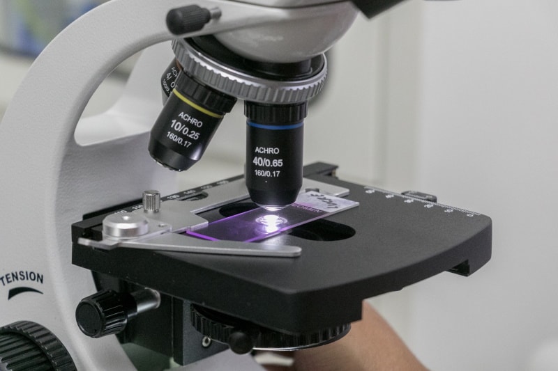
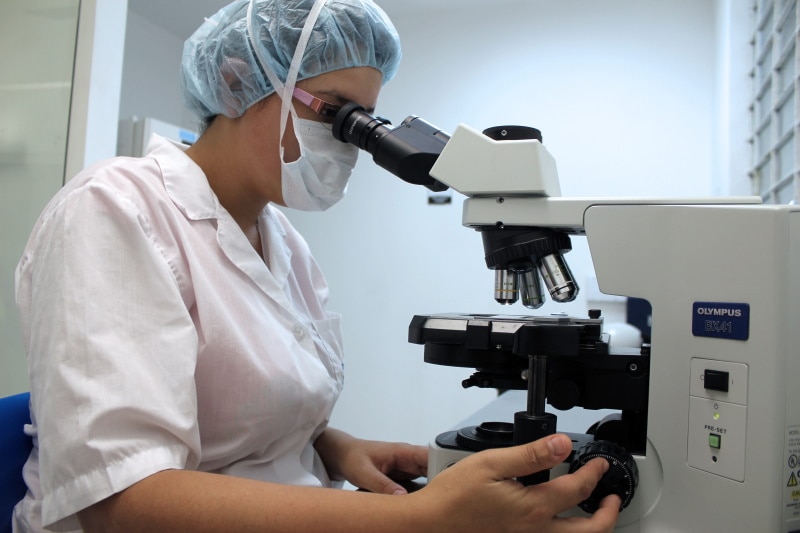
Light Microscopy
It’s the most common type of microscopy used in cell biology. With this type, researchers use visible light to magnify images of cells and cell parts. It has the following applications:
- School and College Labs: When students learn about the basic structure of cells, they use light microscopy because it’s easier to understand.
- Clinical Labs: Light microscopy helps examine blood and other body fluids to look for evidence of infection, disease, or other abnormal conditions.
Brightfield microscopy is the simplest and most common type of light microscopy. It uses light to illuminate the sample and produce an image. Darkfield microscopy is similar to brightfield microscopy, but the background is dark instead of light. That makes it easier to see small, translucent objects like bacteria.
Confocal Microscopy
In this type of microscopy, the focus is kept on one plane while different parts of the specimen are illuminated in succession. A focused laser beam scans the specimen, and the light reflected off it is captured by a photodetector.
You can use this microscopy to study living cells in a tissue. For example, you can study how cells interact with each other or how they change over time.
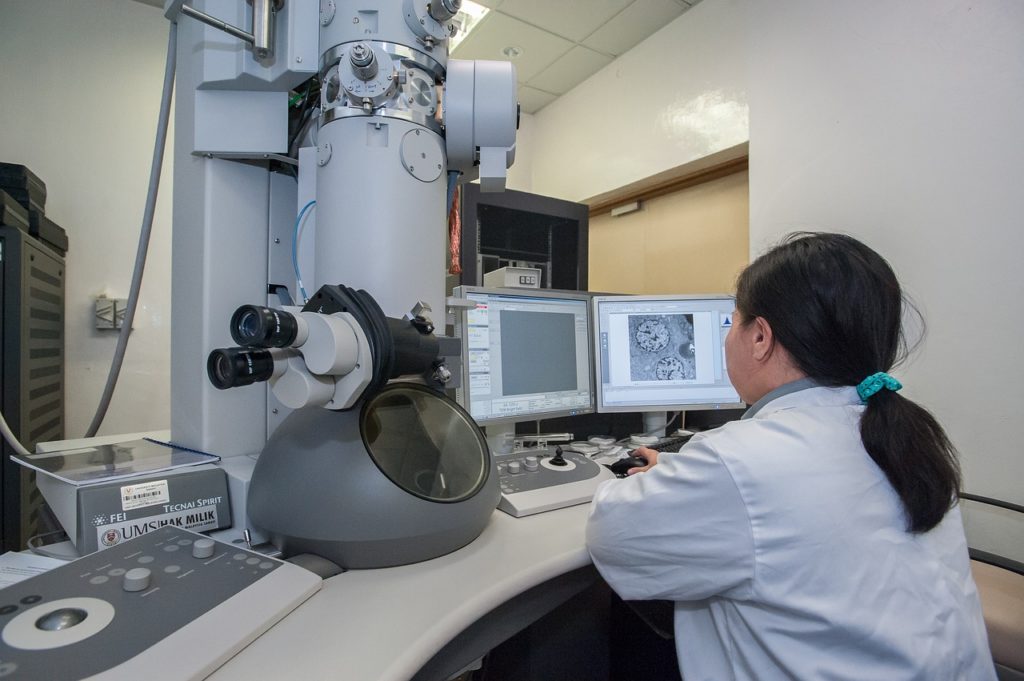
Scanning Electron Microscopy
Researchers who want to study cells’ internal and external structures at a very high level of detail use scanning electron microscopy (SEM).
For example, neuroscience research uses this type of microscopy to study the structure and function of neurons.
Transmission Electron Microscopy
Transmission electron microscopy is another type of microscopy used to study cells at a very high level of detail.
It has similar applications to scanning electron microscopy, but it’s used to study smaller structures, like organelles. For example, researchers might use it to study diseased cells to understand how the disease progresses.
Atomic Force Microscopy
Cells have plenty of attachment points called adhesion sites. These are places where the cell membrane binds to the extracellular matrix or other cells. Likewise, they also have receptors to sense their environment and communicate with other cells.
All these interactions involve physical forces, which you can study using atomic force microscopy (AFM). It uses a cantilever with a sharp tip to map the surface of a cell.
The forces between the tip and the cell surface reveal information about the cell’s adhesion sites, receptors, and other structures.
Importance of Microscopy for Cell Biology
Cell biology studies cells, such as their physiology and structure. A microscope is an essential tool for cell biologists because it allows them to see cells in great detail.
Here are some applications of microscopes in cell biology:
- Study Cell Division: Cell biologists need to study cell division to understand how cells reproduce. The microscope is essential for this because it allows researchers to see the different stages of cell division.
- Observe Cellular Organelles: Microscopes also let cell biologists observe cellular organelles, such as the nucleus, to understand their function and structure.
- Observe Diseased States: Researchers use microscopes to study cells’ surface and internal changes when they’re in a diseased or abnormal state. For instance, a tumor cell looks different than a healthy cell, and microscopes can help scientists see these differences.
Conclusion
Light and electron microscopes can be used to view cells. While the light microscope shows the overall structure of the cell, the electron microscope provides a more detailed internal view. The typical organelles you can view in a cell under a microscope include ribosomes, cell membranes, cell walls, mitochondria, etc.
Cell biologists, researchers, and medical professionals use microscopes to understand cells and their functions better. The standard microscopy techniques they use include brightfield microscopy, phase contrast microscopy, darkfield microscopy, and confocal microscopy.
While each type of microscope has its strengths and weaknesses, they all play an important role in helping us better understand cells.
Featured Image Credit: Piqsels
About the Author Jeff Weishaupt
Jeff is a tech professional by day, writer, and amateur photographer by night. He's had the privilege of leading software teams for startups to the Fortune 100 over the past two decades. He currently works in the data privacy space. Jeff's amateur photography interests started in 2008 when he got his first DSLR camera, the Canon Rebel. Since then, he's taken tens of thousands of photos. His favorite handheld camera these days is his Google Pixel 6 XL. He loves taking photos of nature and his kids. In 2016, he bought his first drone, the Mavic Pro. Taking photos from the air is an amazing perspective, and he loves to take his drone while traveling.
Related Articles:
How to Clean a Refractor Telescope: Step-by-Step Guide
How to Clean a Telescope Eyepiece: Step-by-Step Guide
How to Clean a Rifle Scope: 8 Expert Tips
Monocular vs Telescope: Differences Explained (With Pictures)
What Is a Monocular Used For? 8 Common Functions
How to Clean a Telescope Mirror: 8 Expert Tips
Brightfield vs Phase Contrast Microscopy: The Differences Explained
SkyCamHD Drone Review: Pros, Cons, FAQ, & Verdict