
Optics Mag is reader-supported. When you buy via links on our site, we may earn an affiliate commission at no cost to you. Read more.
What Does Blood Look Like Under a Microscope? (With Pictures)
Last Updated on
Without an optical instrument, you’d assume that blood is a uniformly red liquid, like water mixed with some food coloring.
Your blood will always be red, and the liquid is not just a liquid, as it also has some basic building blocks called cells. If you zoom even further using a microscope, you’ll be able to see other components, such as ions and macromolecules.
Sodium, magnesium, and potassium are all examples of ions found in blood, while the proteins constitute a significant fraction of the total macromolecules. But the question that we’re more interested in answering is, what do all these blood components look like under a microscope?

Plasma
Plasma—sometimes referred to as blood plasma—is the largest component of blood. It’s actually colorless, but under a microscope, it appears straw-colored. That yellowness results from all the dissolved gasses, nutrients, traces of waste, proteins, and more importantly, ions. All these components make up 10% of plasma, while water accounts for 90%.
“What role does plasma play in the human body?”
It serves the following functions:
- Temperature Regulation: We are all warm-blooded creatures. That means we don’t usually rely on external heat sources to regulate our body temperatures. The metabolic processes taking place in our system are the reason why we have the ability to maintain a constant body temperature. Processes that are usually facilitated by plasma.
- Transference: The nutrients drawn from the food that we consume, hormones, electrolytes, and all the essential substances that are requisite for the normal functioning of the body, are usually transported from point A to B using plasma. You’ll also be right to assume that it encourages waste expulsion, seeing as it’s the vehicle that transfers them to the skin, kidneys, lungs, and liver.
- pH Regulation: Did you know our body’s pH cannot be lower than35 or higher than 7.45? If for some reason it falls outside that range, the consequences will be dire. And it’s the different substances found in plasma that always work hard to ensure that doesn’t happen. They act as buffers, hence supporting normal cell functioning.
- Regulating Blood Pressure: When we started, we mentioned that some macronutrients constitute blood—specifically, those that can be found as dissolved substances in plasma. Albumin is part of that community, as it’s classified as a protein. Its principal function is to maintain oncotic pressure, otherwise known as the colloid osmotic pressure. It’s the pressure that pulls fluid (blood) back into your capillaries.
- Offers Immunity: Plasma has immunoglobulins and antibodies, commonly described as disease-fighting proteins. So, without it, various pathogens would take over our bodies.
- Coagulation: Another set of essential proteins found in this component includes factor X, thrombin, and fibrinogen. They are the reason why you don’t bleed out whenever you scrape your knee, as they are meant to encourage clotting.
Red Blood Cells
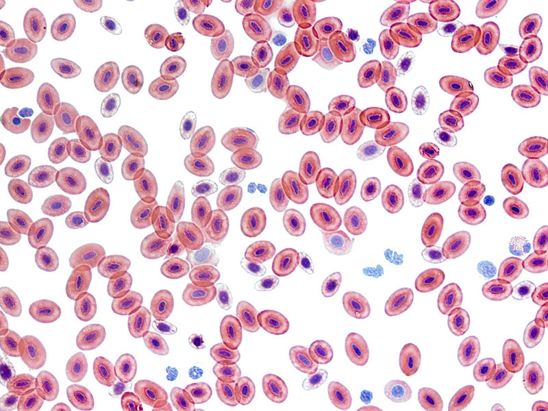
Under the microscope, these cells sort of look like donuts. But donuts without the holes at the center. We like to describe them as biconcave discs, as this shape gives them the ability to squeeze through tiny spaces—especially while carrying luggage meant to be ferried through the capillaries.
By the way, red blood cells don’t usually have the nucleus that you’d typically find in white blood cells. That’s why they can quickly and easily change shape at will and move seamlessly across the semipermeable capillary walls.
This adaptation also increases their surface area to volume ratio, thus ensuring the different gasses effectively diffuse in and out of them. What’s more, the absence of a nucleus means that these cells have more space for hemoglobin, which is a vital iron-containing protein complex that’s relied upon by our bodies in the transportation of oxygen.
Are Red Blood Cells Red in Color?
Yes, they are all red in color because their cytoplasm is rich in hemoglobin.
You need to understand that hemoglobin is essentially a compound that’s made up of globulin chains. And each chain has four protein units, glued together. If you get the chance to examine a mature cell, you’ll learn the hemoglobin molecule always has alpha- and beta-globulin chains—there will be two of each.
If you take a look at the blood smear (a small blood sample studied under a microscope) of an infant or a fetus, you’ll notice that their hemoglobin molecules don’t have any beta-globulin chain, but two gamma chains. That’s because the cell is still in its developmental stage. Gradually, they disappear as the baby grows older.
Each chain comes with a porphyrin compound called heme, and it’s in that heme that you’ll find iron. That iron will bind with oxygen and carbon dioxide, hence facilitating their transportation to the designated areas. Iron is actually what gives our blood its primary red color. If the blood appears bright red, that’s an indication that it’s oxygenated. If it’s dark, it’s deoxygenated.
In case you haven’t caught on already, hemoglobin is what initiates the changes in the red blood cell’s structure. Therefore, if there’s an anomaly affecting how the cell shapeshifts, its functions will be impeded. Ultimately, causing health complications such as sickle cell anemia.
Do Red Blood Cells Have Mitochondria?
No, they don’t. Mitochondria needs oxygen to function, so if the cells had any, they would be forced to use up some of the oxygen being transported to the body tissues. That wouldn’t be efficient in the long run, as the supply would be lower than the demand. Consequently, leading to hypoxia, a condition that has the potential to not only damage the heart, but the brain as well.
Platelets
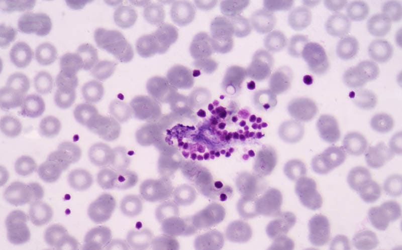
Contrary to popular belief, platelets are not actual cells. They kind of look like cells if you observe them under a microscope, but they are not. We’ve studied these substances long enough to know they are fragments of our body cells. Then again, that doesn’t mean that they are useless, seeing as they have the macro and micronutrients that are vital to the process of blood coagulation.
Unlike cells, platelets have proteins on their surfaces. These proteins are adhesive in nature, making them the perfect remedy for the cracks, or rather breaks, that form on the blood vessel wall. They’ll plug into the wall to cover the breakage, and then ask the body to send more platelets, so that they can stick on the already plugged platelets, to make sure they hold.
The proteins found on platelets are similar to those found on muscle tissue. That’s why they are able to change shape and perfectly fit into the breakage like a piece of a jigsaw puzzle. Under a microscope, they’ll look like a plate, hence the name. But the minute they are signaled to start working, they’ll look like round, extended filaments.
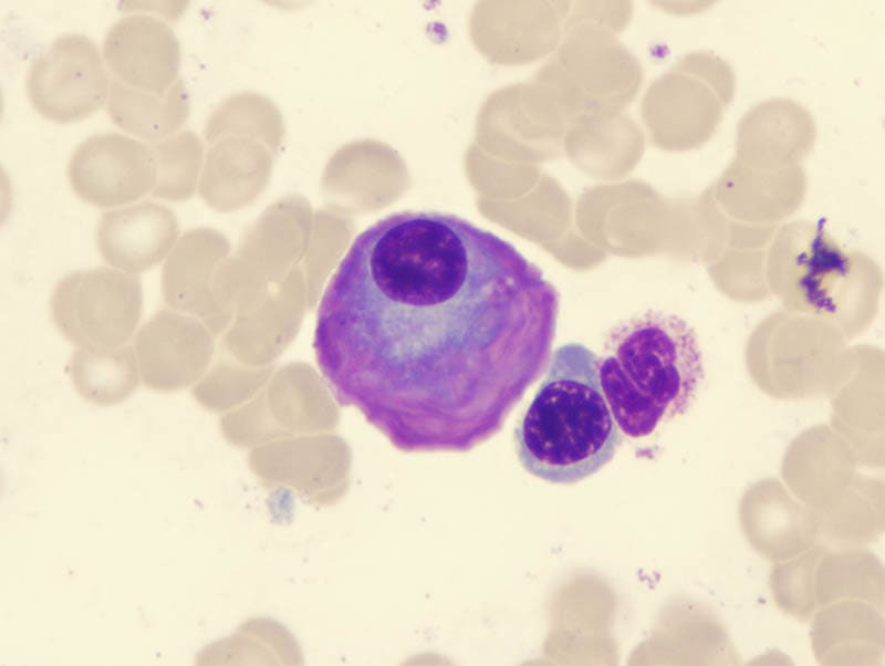
White Blood Cells
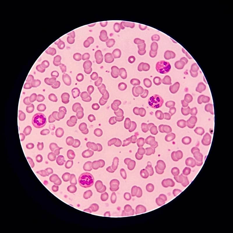
Interestingly, white blood cells aren’t actually white, but colorless. We only call them “white blood cells” because that’s how they often appear whenever we extract a small sample after centrifugation. But if you examine them under the eye of the scope, you’ll realize they are light pink or purplish.
When it comes to their general shape, they are round and have a well-defined center membrane. That membrane represents the boundaries of the nucleus. In addition, these cells are relatively smaller than the red cells.
We forgot to mention that the cells are sometimes called leukocytes. “Leukos” is a Greek word that translates to “white or clear.” This type of cell is not as common as the erythrocytes, as they only account for less than 1% of the total number of cells found in our blood.
White blood cells don’t get themselves involved with the transportation of oxygen in our systems. They are more concerned about invaders such as viruses, parasites, and bacteria that might cause harm if left unchecked. We have five types of leukocyte cells, but they are all grouped into two: agranulocytes and granulocytes.
If you’re able to spot granules in a white blood cell that you’re studying, it’s probably a basophil, eosinophil, or neutrophil cell. Needless to say, they all fall under the granulocyte category. If it doesn’t have any granules, it could be a lymphocyte or monocyte, and is classified as agranulocytes.

Final Thoughts
What does blood look like under a microscope? It looks like many things. Some of its components are colorless, red, pink, purple, weirdly shaped, etc. All that matters is that you’re experienced enough to know what you’re looking at, and how to distinguish it from the rest. Remember, the staining dye that you choose to work with will also influence physical appearance.
See Also: What Do Cancer Cells Look Like Under a Microscope? The Interesting Answer!
Featured Image Credit: TippaPatt, Shutterstock
About the Author Robert Sparks
Robert’s obsession with all things optical started early in life, when his optician father would bring home prototypes for Robert to play with. Nowadays, Robert is dedicated to helping others find the right optics for their needs. His hobbies include astronomy, astrophysics, and model building. Originally from Newark, NJ, he resides in Santa Fe, New Mexico, where the nighttime skies are filled with glittering stars.
Related Articles:
Monocular vs Telescope: Differences Explained (With Pictures)
How to Clean a Refractor Telescope: Step-by-Step Guide
How to Clean a Telescope Eyepiece: Step-by-Step Guide
How to Clean a Rifle Scope: 8 Expert Tips
What Is a Monocular Used For? 8 Common Functions
How to Clean a Telescope Mirror: 8 Expert Tips
Brightfield vs Phase Contrast Microscopy: The Differences Explained
SkyCamHD Drone Review: Pros, Cons, FAQ, & Verdict