
Optics Mag is reader-supported. When you buy via links on our site, we may earn an affiliate commission at no cost to you. Read more.
When Was the Microscope Invented and by Who?
Last Updated on

The first compound microscope was invented sometime around 1590, most likely by Hans and Zacharias Janssen, but there is some debate as to whether it was actually Hans Lippershey, who also filed the first patent for a telescope. Many iterations and versions of the microscope have been invented since then, with Antony Van Leeuwenhoek building the first microscope capable of 270x magnification sometime in the 1670s.
The electron microscope was invented in 1931 by Ernst Ruska, which allowed the viewing of specimens that are smaller than the wavelength of light.
Read on for more details on the early beginnings of this important invention, as well as its many phases of innovation and improvement.

History of the Microscope
Earliest Precursors
The history of the microscope is also the history of optics in general. The first spectacles were made around 1290 in Italy. We don’t know for sure who actually invented them, but the idea of looking through an object to enlarge what is on the other side dates back to Greek and Roman times.
The microscope was born out of an increased understanding of these principles, and the first compound microscope did something that eyeglasses and spectacle makers had never attempted: stack lenses.
The Birth of the Compound Microscope
A compound microscope is so named because it uses more than one magnifying lens to “compound” the amount of magnification possible. There is no certainty as to who actually invented it.
Zacharias Janssen is most widely credited, however, there are conflicting accounts from friends and family. Plus, there is a possibility that the date of the invention was at a time when Zacharias is most likely to have been a small child (his exact date of birth is uncertain), which makes it possible that his next-door neighbor at that time, Hans Lippershey, was the actual inventor.
Regardless of the controversy surrounding the invention of the first compound microscope, it was not a particularly impressive or useful piece of equipment when it was first invented. It was capable of 9x magnification, which is significantly less than handheld magnifying glasses can accomplish today, and it gave a blurry and unfocused image.
But as with all inventions, the potential was realized and over the course of the next hundred years, microscopes would become significantly more powerful and useful.
The Father of Microbiology
Fast-forward to the 1660s, a Dutch fabric merchant named Antony Van Leeuwenhoek was trying to find better ways to closely examine threads and cloth. The compound microscope and the telescope had been around for a while at this point, but Van Leeuwenhoek found his method of magnifying his specimens, which proved to be very effective.
Van Leeuwenhoek’s microscopes used only a single lens, which makes them more akin to modern-day dissecting microscopes, but Van Leeuwenhoek was able to achieve 270x magnification using his single-lens design. His magnification was so high that he was able to discover the existence of bacteria and red blood cells, which earned him the title of “Father of Microbiology”.
His microscopes were so difficult to use, however, that other scientists never adopted them, and it wasn’t until the mid-1800s that compound microscopes evolved enough to achieve similar magnification.
Robert Hooke, a contemporary of Van Leeuwenhoek in England, conducted numerous studies using single-lens microscopes available at the time and published the Micrographia. This publication documented all the things he had observed, which included his own drawings of what he saw. It was in the Micrographia that Hooke coined the term ‘cell’ to describe the basic unit of life.
The Evolution of Microscopes
Throughout the centuries, microscopes have continued to be innovated and improved. The compound microscope continued to evolve and provide higher magnification, but the images seen through them suffered significant chromatic aberration and distortion, which made the images not as useful as they could otherwise be.
Nearly a hundred years after Van Leeuwenhoek, Chester Moore Hall invented the achromatic lens, which resolves the chromatic aberration in compound microscopes by specifying that the second lens must be of a different shape than the first lens. By doing this, he could realign the colors that were split apart by the lens providing the magnification.
Nearly a hundred years after that, Joseph Lister was finally able to resolve the distortion problem. Even in many modern-day magnification devices, the image viewed distorts as it gets closer to the edge of the lens. This is called spherical aberration and happens because the angle of the light exiting the lens changes based on what part of the lens it is passing through.
From that point, microscopic innovations leaped forward as trial-and-error was replaced with mathematics, and light microscopes quickly became so advanced that they approached the theoretical magnification limits of the wavelengths of visible light.
Beyond Visible Light
The UV microscope was first introduced by a company called Zeiss and was able to provide magnification that far exceeded that of visible light.
Shortly after, Frits Zernike, a Dutch physicist, invented the phase-contrast microscope, which essentially detects variations of light amplitude to give minute details about the specimen. The phase-contrast microscope worked well for examining specimens that were still alive without the need to stain them as would be required in a compound microscope.
Right around the same time, the first electron microscope was invented by Ernst Ruska and Max Knoll. Electron microscopes use a beam of electrons the same way that a light microscope uses a beam of light to illuminate the specimen. Since electrons have a wavelength far smaller than visible or UV light, electron microscopes can achieve magnification far beyond that of a traditional light microscope.
Today, electron microscopes come in two varieties: a transmission electron microscope that sends the beam of electrons through the specimen and into the microscope, and a scanning electron microscope that bounces the beam of electrons off the specimen and back up into the lens. These modern electron microscopes are capable of up to 2 million times magnification.
Conclusion
The idea of using an object to magnify things around us has been available for thousands of years. The first major landmark in the development of microscopes was the invention of the first compound microscope in the late 1500s, but microscopes did not begin to be truly useful until much later as single-lens microscopes became more advanced in the 1600s.
As the centuries moved past, so too did the evolution of the microscope. Image quality improved, magnification increased, and eventually, microscopes moved past the limits of light itself to show us things on a subatomic level.
See also:
- Transmission (TEM) vs Scanning (SEM) Electron Microscopes: What’s the Difference?
- Brightfield vs Phase Contrast Microscopy: The Differences Explained
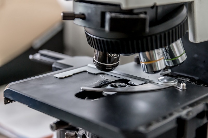
Featured Image Credit: Artem Podrez, Pexels
About the Author Robert Sparks
Robert’s obsession with all things optical started early in life, when his optician father would bring home prototypes for Robert to play with. Nowadays, Robert is dedicated to helping others find the right optics for their needs. His hobbies include astronomy, astrophysics, and model building. Originally from Newark, NJ, he resides in Santa Fe, New Mexico, where the nighttime skies are filled with glittering stars.
Related Articles:
When Were Binoculars Invented? History, Today & Future
How to Clean a Refractor Telescope: Step-by-Step Guide
How to Clean a Telescope Eyepiece: Step-by-Step Guide
How to Clean a Rifle Scope: 8 Expert Tips
Monocular vs Telescope: Differences Explained (With Pictures)
What Is a Monocular Used For? 8 Common Functions
How to Clean a Telescope Mirror: 8 Expert Tips
Brightfield vs Phase Contrast Microscopy: The Differences Explained