
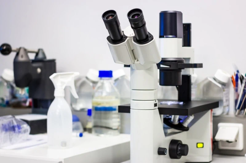
Optics Mag is reader-supported. When you buy via links on our site, we may earn an affiliate commission at no cost to you. Read more.
What Is a Phase Contrast Microscope? The Interesting Answer!
Last Updated on
It’s possible to study, examine, and observe objects—or parts of those objects—that are too tiny to be seen with the naked eye. That whole process is called microscopy because you’ll be using an optical instrument called a microscope.
We have different types of microscopes, and the phase-contrast microscope is one of them. Once you’re done utilizing it, the process will now be described as phase-contrast microscopy. A phase-contrast microscope turns the phase shifts in light passing through a specimen into brightness changes in the image.

How Does a Phase-Contrast Microscope Work?
Living cells (specimens) that are unstained are incapable of absorbing sufficient light. As a result, if you look at the images generated, you’ll note that there’s an insignificant difference in their intensity distribution. In other words, the specimens are barely visible when examined using a brightfield microscope.
That’s where the phase-contrast microscope comes in. This instrument has the power to transform the phase shifts (that’s the disparity between the phases of two periodic signals) in the light being transmitted through a transparent object, to the brightness changes observed in an image.
What’s a Phase Object?
In this field, any unstained specimen that can’t absorb light is referred to as a phase object. And they always experience difficulties absorbing the light being transmitted through them because they keep changing the phase of light that they diffract. Once you’ve done the math, you’ll learn that the phase-shift difference between the background light and the one diffracted is about ¼ wavelength.
Even though that’s a slight phase difference, the human eye wasn’t designed to detect that kind of variation. We are only capable of detecting variations in light intensity and frequency. Hence, the reason why it’s difficult to interpret the changes in the images produced by a brightfield microscope when observing an unstained specimen.
What is Phase Contrast?
Phase contrast is the technique that allows the generation of high contrast images of a phase object, by widening the light phase difference. So, put simply, it’s the process that separates our specimen’s diffracted light from the background light.
Just before the image produced hits the plane where it’s supposed to be projected, the microscope’s phase plate will advance or slow down the background light by ¼ of a wavelength. This action causes a constructive or destructive interference, ultimately resulting in changes in the brightness level of the areas we couldn’t see in the image.
Types of Phase Contrast Microscope
The type of phase contract instrument that you have will depend on its phase plate design. There are several, but the most common types are the positive and negative phase contrast.
Positive Phase Contrast
Most microscopes found on the market have this type of phase contrast. When examining the thicker organelles in a living organism’s cell using this scope, the first thing that you’ll notice is that they appear unusually darker against a light background.
And that’s ultimately the goal of this type of scope. It advances the direct light’s wave by ¼, to initiate deconstructive interference that leads to darker details on a light background.
It’s ideal for researchers looking to examine aquatic microorganisms, particularly those who are more interested in studying a cell’s mitochondria or nucleus.
Negative Phase Contrast
The negative contrast does the complete opposite. It makes those thicker parts of a cell appear brighter when contrasted against a dark background. The direct light in this instance will be slowed down by ¼ wave, to generate constructive interference, which results in brighter details against a dark background.
Negative phase contrasts are suitable for scientists who often examine protists. Protists are described as eukaryotic living organisms that are neither classified as animals nor plants.
Inverted Phase Contrast
This type of microscope is very rare. It’s different in that it operates like a brightfield microscope, but with phase contrast capabilities. The phase plates are normally located outside the objective lens, making sure they are able to generate a phase contrast.

What Are the Parts of a Phase-Contrast Microscope?

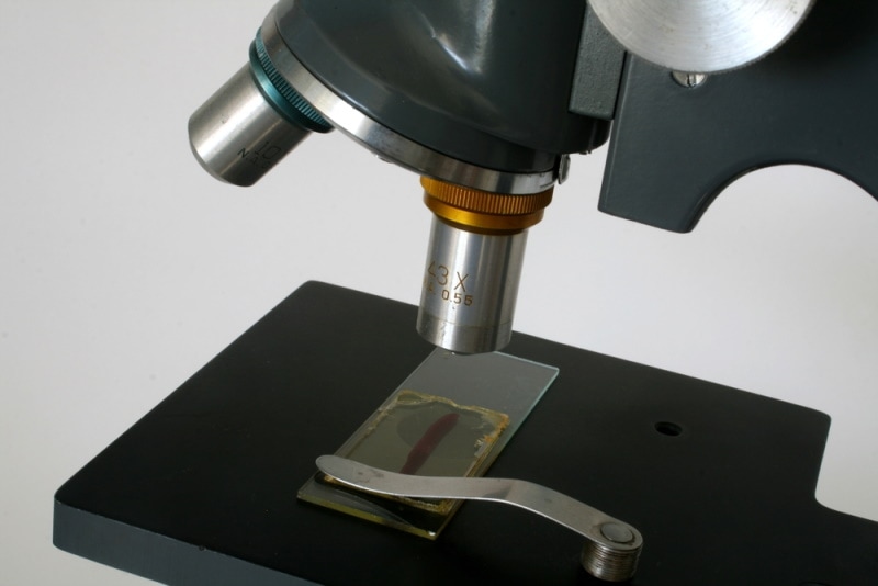
This microscope is an iteration of a simple microscope, but with a few modifications. That means you’ll find all the usual components, including an objective lens, arm, base, specimen stage, condenser lens, and more importantly, the eyepiece (ocular lens).
The only parts that make it different are the phase plate and the annular diaphragm. Some sources will call the latter a phase annular or annular ring.
Annular Diaphragm
The diaphragm has two parts: a circular annular groove and a disc. The function of the groove is to transmit the light rays to the object. This groove will be located under the microscope’s condenser lens.
Phase Plate
The plate is always transparent and takes the form of a disc. You’ll also notice a ring on it, which is filled with material often described as phase advancing and retarding matter. That ring is sometimes lighter than other sections of the plate, or darker. Basically, the addition of the material influences the light amplitude.
The annulus diaphragm and the phase plate have to be positioned the same way, as they usually work as a unit. And what that means is that it will also be part of the objective lens system, in addition to having its own complementary and conjugate components.
Where Is It Used?
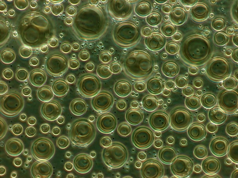
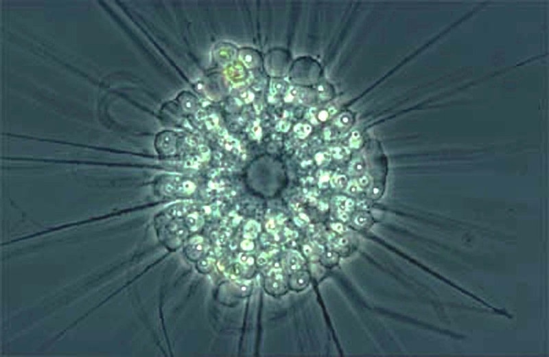
You can use a phase-contrast microscope to study anything that qualifies as a specimen and is transparent. For example, you could use it when examining fibers, microorganisms, glass fragments, thin tissue slices, latex dispersions, subcellular particles, and even lithographic patterns.
It’s also applicable in situations where an expert is trying to detect bacterial elements, such as endospore, on a surface.
Advantages of Phase Contrast Microscopy
- Examining thin slices of specimens would be very difficult if we only had compound or simple microscopes. So, we have the phase-contrast scope to thank for that.
- This tool can produce high-resolution images of the intercellular parts of any living organism’s cell.
- Users don’t have to worry about stains reacting with their specimens, seeing as the phase contrast microscope doesn’t require staining.
- Irrespective of how transparent your specimen is, the image will always be very visible.
- In addition to creating images, this microscope can also record videos of the specimen being observed.
- The contrast of the images generated is usually high.
- It allows the user to examine a living cell in its natural state, without having to be fixed or killed.
Disadvantages Of Phase Contrast Microscopy
- There’s a possibility that you’ll have to deal with halo or shade-off effects while examining your specimens.
- The photomicrography is not all that great if you use green or white lights. The images produced will either appear green or gray.
- Not ideal for thick specimens, since the images are always distorted.
- Assembling the different components of the scopes makes the whole examination process costlier.
Frequently Asked Questions
What’s The Principle Behind a Phase-Contrast Microscope?
To better understand this concept, you have to see light as a wave. Now we have a source, and the beams of light emanating from our source, are being released in synchronization. When the first beam hits a specimen, it does so with a relatively higher refractive index, compared to that of air. The beam is then compelled to slow down but increase its waves in proportion to the specimen’s thickness and refractive index.
Then we have the second beam that doesn’t come into contact with any specimen. It only travels through air, and that’s it. The two beams will meet at some point, but this time round, there won’t be synchrony. We’ll have what’s called a light phase difference.
You mustn’t forget the fact that all types of specimens can diffract light. And when you see an image being produced, it’s because there was a phase difference between the direct light and the one that’s been diffracted. The thing that essentially gives the stained specimen an edge over the unstained object is the adequacy of the phase difference—a wider phase difference simply translates to a clearer image.
Who Invented Phase Contrast?
Frits Zernike, who was a physicist in 1932, was the one who found a remedy to this problem. He’s the one who first discovered that the unstained specimens generated terrible images due to an inadequate phase difference between the direct and diffracted light. So, to separate the light rays, he added a disk in the condenser.
When he realized the images were still not up to par, he added a special kind of plate at the back of the objective lens’ focal plane. And before he knew it, he had achieved something that would go on to win him a Nobel Prize. The disk is now what’s referred to as the annular ring, and the special plate is the phase plate.
What is the maximum magnification of a phase contrast microscope?
The magnification of a phase contrast microscope depends on the microscope itself, although most include removable annular diaphragms that can be replaced with those of different magnification levels. Generally, the greatest magnification level of this type of microscope is 100x.
How can you tell if a microscope is phase contrast?
Without looking through a microscope, it can be difficult to tell if it is phase contrast or not. When looking at objects under a microscope, if there are halos around the objects or there is blurring, it is likely a phase contrast microscope and not a widefield one.
Can phase contrast be used with stained samples?
Phase contrast can be combined with staining and other methods of microscopy. Doing so can enhance the visualization of the subject being viewed, and combining these two microscopy techniques can yield highly detailed results.
Final Thoughts
The phase-contrast microscope has a light path that’s way different from the one found in a simple or compound scope. It’s not only very efficient but also effective at producing high-resolution images. Like several other instruments, it comes with a set of merits and demerits. But the good news is that the perks outweigh the weaknesses.
Featured Image Credit: Catalin Rusnac, Shutterstock
About the Author Robert Sparks
Robert’s obsession with all things optical started early in life, when his optician father would bring home prototypes for Robert to play with. Nowadays, Robert is dedicated to helping others find the right optics for their needs. His hobbies include astronomy, astrophysics, and model building. Originally from Newark, NJ, he resides in Santa Fe, New Mexico, where the nighttime skies are filled with glittering stars.
Related Articles:
What Is the Best Binocular Magnification for Hunting? Optical Features Explained
How to Clean a Refractor Telescope: Step-by-Step Guide
How to Clean a Telescope Eyepiece: Step-by-Step Guide
How to Clean a Rifle Scope: 8 Expert Tips
Monocular vs Telescope: Differences Explained (With Pictures)
What Is a Monocular Used For? 8 Common Functions
How to Clean a Telescope Mirror: 8 Expert Tips
Brightfield vs Phase Contrast Microscopy: The Differences Explained