
Optics Mag is reader-supported. When you buy via links on our site, we may earn an affiliate commission at no cost to you. Read more.
What Is the Ocular Lens on a Microscope? Interesting Facts
Last Updated on

The ocular lens is the lens you look through when using a microscope. It is also known as the eyepiece. The ocular lens is typically made of glass and is the last lens in the microscope’s optical path before your eye. Therefore, it magnifies the image that is being focused on by the objective lenses.
Most ocular lenses have a magnification of 10x, but some microscopes have ocular lenses with a lower or higher magnification. In this article, we explain the function, types, and benefits of the ocular lens in detail.

How Does It Work?
The ocular lens on a microscope works by bending the light that enters it. That’s because the lens is curved, and when light hits a curved surface, it bends.
The amount the light bends depends on how curved the surface is. The ocular lens is usually convex, which means that it curves outward. Here’s the mechanism of action of a microscope’s ocular lens:
- The viewer looks through the ocular lens, which is mounted on an eyepiece at the top of the microscope.
- Light enters the microscope through an object lens located below the stage (the platform where the specimen is placed).
- The light then passes through the objective lens and hits the ocular lens.
- The ocular lens bends the light and magnifies the image of the specimen.
- The viewer sees a magnified image of the specimen through the ocular lens.
All microscopes have at least one ocular lens, and some have two. You can adjust the magnification amount by changing the objective lens’s power. The ocular lens just magnifies the image already magnified by the objective lens.

Types of Ocular Lenses
There are a few different types of ocular lenses used in various microscopes. The common ones are as follows.
Wide-Field Lens
A wide-field lens on a microscope provides a large field of view, allowing the user to quickly take in a great deal of information. Such a lens is especially useful for scanning an area for potential targets or abnormalities.
Super-Field Lens
A super-field lens is used in stereoscopic microscopes, providing a large field of view to observe fast-moving objects.
The lenses are composed of two glass plates with a liquid filling in between, and the distance between the plates can be adjusted to change the magnification.
Super-field lenses are used in various applications, including medical research, industrial inspection, and security surveillance.
Compensation Lens
A compensation lens is an ocular lens that helps correct the objective lens’s aberration. For example, in the case of a spherocylindrical lens, it is used to bring back the image in focus by canceling out the spherical aberration.

Periplan Lens
A Periplan lens in a microscope can correct chromatic aberration while providing high magnification.
It is a compound lens consisting of a convex lens made of glass with a refractive index lower than the surrounding medium and a concave lens made of a material with a refractive index higher than that of the surrounding medium.
Chromatic aberration is a type of distortion that occurs when light waves of different wavelengths are focused at various points on the lens. It is common in simple lenses, such as those found in microscopes.
Thus, a Periplan lens is helpful in applications where the viewer cannot afford chromatic aberration to distort the image.
Huygen Lens
A Huygen lens has two plano-convex lenses, with their convex sides facing each other. It forms a Huygenian eyepiece.
The Huygen lens forms a real image, which is the same size as the object. Therefore, the Huygen lens has a short focal length and a long back focal length.
Ramsden Lens
The distinct characteristic of the Ramsden lens is that it has a field stop on the outer side of the lens tube. The field stop allows the user to see a small 2mm central circle of the field of view and completely cuts off the outer ring of light.
It results in an increased perception of sharpness and brightness in the central area of the field of view.

Where Is the Ocular Lens Used?
An ocular lens is one of the essential parts of a microscope since it is responsible for gathering light and forming the image the user sees. It is the lens closest to the eye and is usually made of a material with a high refractive index, such as glass. The ocular lens is placed at a distance from the specimen equal to its focal length. It ensures optimal magnification of the image the user is viewing.
Anatomy of an Ocular Lens
An ocular or eyepiece lens on a microscope has several internal and external parts. Here’s a breakdown of the lens’s anatomy.
- Rubber Eyecup: It is the black rubber ring that sits on the outside of the ocular lens. Its primary purpose is to provide comfort and block out external light when pressed against the eye.
- Field Lens: The field lens is the first glass lens that light passes through after entering the ocular. It creates a real image of the object being viewed by the user.
- Diopter Adjustment: The ocular lens has a diopter adjustment, a knob on the side of the lens that can be turned to focus the image.
- Eyetube Mounting Flange: The eyetube mounting flange is the portion of the lens that attaches to the microscope’s eyetube.
- Aperture: The aperture is the small opening in the center of the ocular lens that allows light to pass through.
- Eyetube Locator Tab: It’s on the side of some ocular lenses and is used to align the lens with the microscope’s eyetube.
- Lens Triplet: Ocular lenses also have a lens triplet, a group of three lenses that work together to create a clear image.
- Lens Doublet: The lens doublet is located above the triplet. It also helps create a clear image by bending the light that passes through it.
- Coverslip: The coverslip is a piece of glass that sits on top of the lens and protects it from damage.

What Do the Inscriptions on a Microscope’s Ocular Lens Indicate?
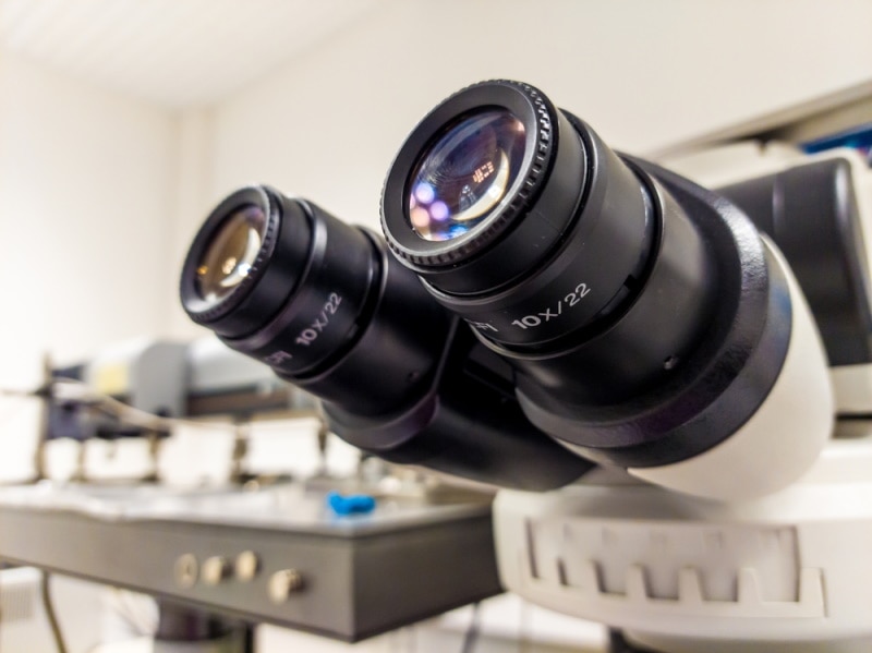
The ocular lenses, or eyepieces, of a microscope, have numbers inscribed on them that indicate the power of the lens. These numbers are usually in the range of 4X to 100X.
The number on the right ocular lens is always 10X greater than the number on the left ocular lens. So, for example, if the left ocular lens is inscribed with the number “40,” the right lens will be inscribed with the number “100.”
Apart from the numbers, you’ll also see letters inscribed on the eyepiece. These include UW, WF, SW, HE, CF, and UWF. Here’s what they mean:
| WF | Widefield |
| UWF | Ultra-wide field |
| SW | Super-widefield |
| HE | High eyepoint |
| CF | Corrected for astigmatism |
Some microscopes have two ocular lenses (eyepieces) that can be used interchangeably. You’ll find the magnification power inscribed on both the ocular lenses in such microscopes.
Compensation microscopes have inscriptions, such as K, comp, or C. You’ll also see the magnification next to these letters.
What to Consider When Choosing an Eyepiece Lens for a Microscope
It’s best to buy an objective lens and then get an eyepiece lens that will fit it. For example, if you’re using a compound microscope, you’ll need to determine the magnification of your objective lenses and purchase an eyepiece lens that will work with those objective lenses.
Some factors you’ll need to consider when choosing an ocular lens include:
Field of View
It is the diameter of the area that you can see through the microscope. A larger field of view allows you to see more area at once, while a smaller field of view means you’ll need to scan back and forth to take in the entire area you’re observing.
Magnification
The magnification is the number printed on the side of the eyepiece lens and tells you how much larger the image will appear when using that lens.
For example, a 10x eyepiece lens will make the image appear 10 times larger than it would without the lens. Select the magnification based on the applications you need the microscope for.
Focal Length
The distance from the center of the lens to the point where the image is in focus is the lens’s focal length. A shorter focal length keeps the image in focus at a closer distance, while a longer focal length is best for viewing objects further away.
Diopter Adjustment
Most eyepiece lenses have a diopter adjustment, which is a way to fine-tune the focus for your eyesight. Again, you should consider your needs when selecting a suitable adjustment range.
Besides these factors, you might have to consider your budget, brand, warranty, and other factors. If you already have an objective lens, the key is only to buy the eyepiece the objective lens’s manufacturers recommend using with their lenses.

Advantages of Ocular Lens
An ocular lens on a microscope has many advantages. Some of them are:
- Field of View: The ocular lens provides a large field of view. You can see more regions of the sample with an ocular lens than with other lenses.
- Depth of Field: The ocular lens also provides a considerable depth of field. It means you can see far-away and close-up objects with the same lens.
- Adjustable: Since the ocular lens is adjustable, you can change its magnification. You can use a lower magnification to see a larger area of the sample or a higher magnification to see a smaller area in more detail.
- Corrects Aberrations: Some types of ocular lenses can correct chromatic aberration caused by different light colors bending at different angles.
- Image Clarity: You can see a clear image with an ocular lens because it forms a real image on the retina of your eye. A “real” image means that the image is upside down and left to right reversed.
Disadvantages of Ocular Lens
An ocular lens also has some disadvantages. Some of them are:
- Blurring: The lenses in the eye are not perfect as they may cause the image to be blurred. The ocular lens can magnify this blur.
- Lens Flares: If you look at a bright light with an ocular lens, you may see streaks of light called lens flares.
- Ghost Images: Sometimes, you may see a faint image of the original object next to the real image. It is called a ‘ghost image’ and can be due to the reflections off the surface of the lens.
- Eye Strain: Looking through an ocular lens for a long time can cause eye strain.
How to Care for an Ocular Lens: Tips for Microscope Users
Since the ocular lens is the lens you look through when using a microscope, it is crucial to keep it clean. The following tips will help you care for your ocular lens and ensure that it stays in perfect condition for a long time.

Clean Regularly
Clean the ocular lens regularly with a soft, dry cloth. If there is any dirt or debris on the lens, use a slightly dampened cloth to wipe it away gently.
Be careful not to get any water on the lens, as this can damage it. Also, when moving the microscope from one place to another, keep the ocular lens covered so that it doesn’t come into contact with anything that could potentially scratch or damage it.
Avoid Touching
It is essential to avoid touching the ocular lens with your fingers, as the oils from your skin can transfer to the lens and cause streaks. If you must touch the lens, do so with a clean, dry cloth.
Store Properly
When not in use, store the microscope in a clean, dry place. If possible, keep it in a dust-free environment. If you have no other choice but to keep the microscope in a dusty cabinet, cover the ocular lens with a clean cloth to protect it.
Replace When Needed
Since the eyepiece lens is interchangeable, you can replace it with a new one if it becomes scratched or damaged. Get the correct size lens for your microscope, as using an incorrect size lens can damage the microscope.
You should consult the microscope’s manufacturers or a qualified technician to find out what size lens you need.

Frequently Asked Questions
Is the Ocular Lens Same as the Eyepiece?
Yes, the ocular lens is another name for the eyepiece. The ocular lens is the lens you look through when using a microscope.
What Is the Standard Magnification of an Ocular Lens?
Most ocular lenses have a magnification of 10x. It means that the image you see through the lens will be ten times larger than it would be with the naked eye.
Can You Use a Higher-Powered Ocular Lens on a Lower-Powered Microscope?
Using a higher-powered ocular lens on a lower-powered microscope is not recommended. The image will not be in focus and will be difficult to see.
What Is the Function of the Eyepiece Lens?
The eyepiece lens magnifies the image that is produced by the objective lenses. Thus, if you are using a 10x eyepiece lens, the image will be magnified ten times after an objective lens has magnified it.
What Is a Field Stop?
The ocular lens has a field stop mechanism that limits the amount of light that enters the eye. As a result, it allows you to see a clear image by blocking the surrounding light. For instance, if you are looking at a white object, the field stop will help you see it more clearly by blocking the background light.
How Do You Use the Ocular Lens?
Firstly, you need to make sure that the ocular lens is clean. If it is dirty, it will affect the quality of the image. Next, adjust the microscope so that the image is in focus. You can do this by moving the stage up and down. Finally, look through the ocular lens and focus on the image.
Conclusion
The ocular lens on a microscope is the lens you look through to see the specimen. It is usually one of the two lenses on a compound microscope. The other lens, below the ocular lens, is the objective lens. The ocular lens is also called the eyepiece.
The ocular lens on a microscope works by bending (refracting) light rays that pass through it. As a result, it causes the specimen to appear magnified. The amount of magnification depends on the power (focal length) of the ocular lens.
Featured Image Credit: Piqsels
Table of Contents
- How Does It Work?
- Types of Ocular Lenses
- Where Is the Ocular Lens Used?
- Anatomy of an Ocular Lens
- What Do the Inscriptions on a Microscope’s Ocular Lens Indicate?
- What to Consider When Choosing an Eyepiece Lens for a Microscope
- Advantages of Ocular Lens
- Disadvantages of Ocular Lens
- How to Care for an Ocular Lens: Tips for Microscope Users
- Frequently Asked Questions
- Conclusion
About the Author Jeff Weishaupt
Jeff is a tech professional by day, writer, and amateur photographer by night. He's had the privilege of leading software teams for startups to the Fortune 100 over the past two decades. He currently works in the data privacy space. Jeff's amateur photography interests started in 2008 when he got his first DSLR camera, the Canon Rebel. Since then, he's taken tens of thousands of photos. His favorite handheld camera these days is his Google Pixel 6 XL. He loves taking photos of nature and his kids. In 2016, he bought his first drone, the Mavic Pro. Taking photos from the air is an amazing perspective, and he loves to take his drone while traveling.
Related Articles:
Binocular Magnification Chart: Numbers & Distances Compared
What Is the Best Binocular Magnification for Hunting? Optical Features Explained
When Were Binoculars Invented? History, Today & Future
How to Clean a Refractor Telescope: Step-by-Step Guide
How to Clean a Telescope Eyepiece: Step-by-Step Guide
How to Clean a Rifle Scope: 8 Expert Tips
Monocular vs Telescope: Differences Explained (With Pictures)
What Is a Monocular Used For? 8 Common Functions