
Optics Mag is reader-supported. When you buy via links on our site, we may earn an affiliate commission at no cost to you. Read more.
What is Dark Field Microscopy Used For? The Surprising Answer
Last Updated on

Ancient peoples knew of the concept of microscopy from simple observation. Roman philosopher Seneca wrote of writing appearing larger when viewed through a water-filled glass. However, the inventor is unknown, although many individuals created variations, beginning in the 13th century with English philosopher Roger Bacon.
The 17th century was a time of rapid development from other inventors, such as Antonie van Leeuwenhoek. These works culminated in the brightfield microscope, which you undoubtedly used in school. It uses light focused on a specimen during magnification using either a mirror or other powered source. However, the invention has continued to evolve, with the dark field microscope opening up another research avenue. It steps up to the plate when the brightfield pales in comparison.

How Does It Work?
To understand dark field microscopy, we must begin with brightfield microscopy. You look through the ocular lens, viewing what the objective lens has magnified. The specimen is on a prepared slide with a cover slip on top, sitting on the stage. A light source sits on the base of the microscope, shining into the condenser on the bottom of the stage. It does just what its name implies, illuminating the slide from the bottom.
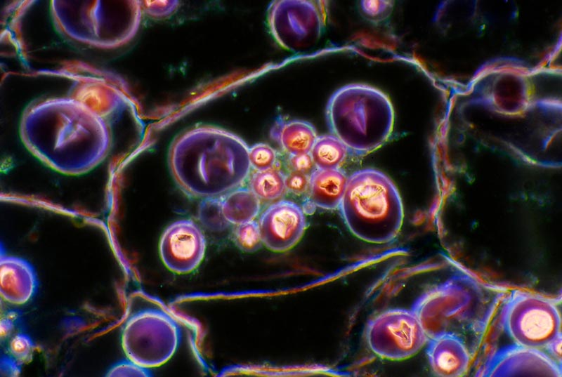
Brightfield microscopy uses a full cone of light. Dark field microscopy differs with its hollow cone. The result is light with various angles, illuminating the specimen but not the area around it. It may seem like a technicality, but the changes in what you see through the ocular lens are profound. You can think of it as a riff on brightfield microscopy that broadens its usefulness and value with specific applications.
When Is It Used?
Sometimes, brightfield microscopy doesn’t deliver desirable results. That typically occurs when the refractive indices or contrast isn’t enough to provide sufficient detail. You may not be able to see anything but a simple shape. Some microorganisms, like amoebas, are transparent. You need the optimal contrast to see the organelles within it. That’s where dark field microscopy comes into play.
It provides the necessary contrast to see specimens in more detail. Scientists and the healthcare industry use it to view algae, bacteria, and blood cells. It’s an excellent alternative for observing live specimens where a stain might kill them. That’s not to say it replaces this process. It means you can get by with less, making it easier to study your specimens.
However, its applications aren’t limited to microbiology. Scientists also use dark field microscopy to detect flaws or cracks in metals that aren’t visible to the naked eye. It’s also valuable for looking at diamonds and other gems to determine their quality and grade. Dark field microscopy isn’t a new technique. Instead, it’s a variation on the theme that extends the usability of the technology.
Advantages of Dark Field Microscopy
The other alternative to using brightfield microscopy is phase contrast microscopy. Unfortunately, it’s an expensive technology. Value-priced devices are a poor substitute for dark field ones. It’s affordable with the addition of an occulting disk. That’s considerably cheaper than buying another device for these applications. Researchers appreciate it since it is less likely to harm live specimens due to stains. That makes it priceless for some fields.

Disadvantages of Dark Field Microscopy
However, dark field microscopy isn’t without its disadvantages. It can kill live specimens not because of the stains, but from the focused light on the field of view. That limits how you can use it, at least in biology. These same disadvantages aren’t a factor with metals or gemstones. Therefore, it boils down to how you want to use dark field microscopy, taking into account its risks.

Frequently Asked Questions (FAQ)
How can I make a dark field microscope?
We mentioned the occulting disk you can use to convert a brightfield model to this type. The trick is all about its placement. You need to put it between the condenser and the light source to change its focus.
What do specimens using this microscopy type look like?
The difference lies in the contrast. You’ll go from looking at a gray blob with specs to a high-contrast specimen with intricate detail. The results are often dramatic compared to the brightfield way.
Who invented dark field microscopy?
Amateur British scientist Joseph Jackson Lister invented the first device using this technology in 1830. His interest in natural history propelled his development of dark field microscopy, which may explain its present-day applications.

Final Thoughts
Dark field microscopy opens up worlds that brightfield microscopy can’t see. It builds on the principles of the technology to make it more useful across a broader range of fields. It doesn’t detract from the magnifying properties of other devices. Instead, its focus is on improving the contrast within your specimens. You can think of it as viewing the microscopic world through a different lens.
Featured Image Credit: Mike Rosecope, Shutterstock
About the Author Chris Dinesen Rogers
Chris has been writing since 2009 on a variety of topics. Her motto with all of her writing is “science-based writing nurtured by education and critical thinking.” Chris specializes in science topics and has a special love for health and environmental topics, and animals of all shapes and sizes.
Related Articles:
What Is the Best Binocular Magnification for Hunting? Optical Features Explained
How to Clean a Refractor Telescope: Step-by-Step Guide
How to Clean a Telescope Eyepiece: Step-by-Step Guide
How to Clean a Rifle Scope: 8 Expert Tips
Monocular vs Telescope: Differences Explained (With Pictures)
What Is a Monocular Used For? 8 Common Functions
How to Clean a Telescope Mirror: 8 Expert Tips
Brightfield vs Phase Contrast Microscopy: The Differences Explained