What Is a Condenser on a Microscope (And What Does It Do)
Last Updated on

A microscope typically consists of the following parts: an eyepiece lens, an objective lens, a stage, a base, a condenser, and an illuminator. All these parts have their specific functions that ensure the microscope works correctly.
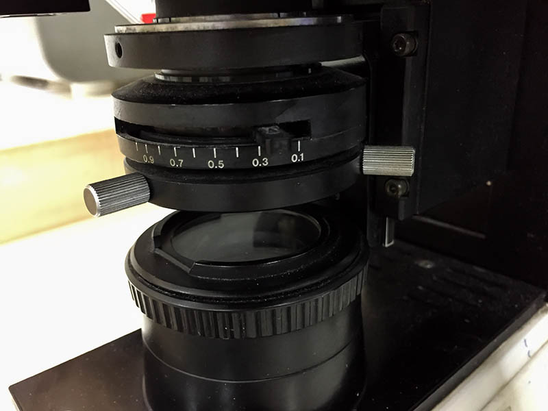
Among these, the condenser is one of the most critical components as its responsibility is to gather light and reflect it through the objective lens. It results in a brightly lit specimen on the stage that you can see through the lens.
Let’s magnify (pun intended) the role of a condenser and discuss the different types of microscopes you can expect to find on a microscope.

What Is a Condenser on a Microscope?
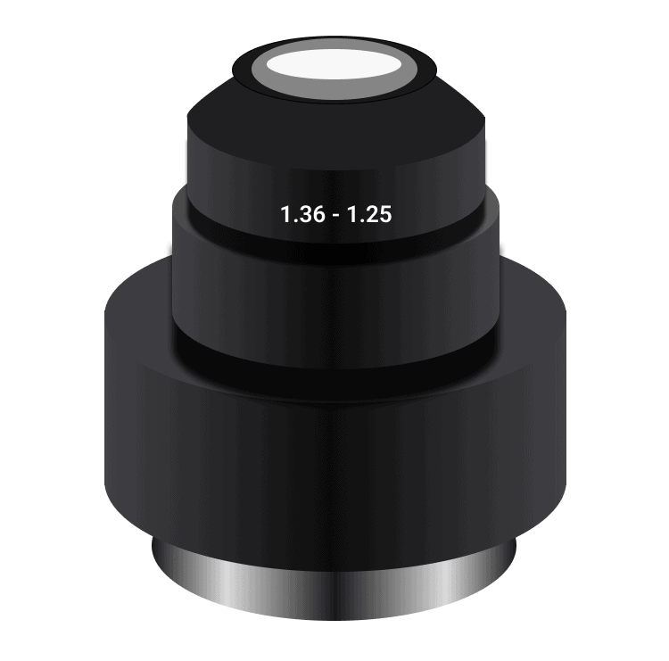
A condenser is a lens or system of lenses used to converge or diverge light. In microscopy, a condenser is a necessary component that provides illumination to the sample you’re viewing.
The most common condenser type is the Abbe condenser, which uses a combination of lenses to control the path of light as it enters the objective microscope lens. Since high-power objective lenses have tiny diameters, they need ample light to produce a clear image. Therefore, the light must be concentrated and directed to pass through the center of the objective lens. The condenser is responsible for this task.
Condensers provide bright-field illumination, which is the most common type of microscopy. In bright-field microscopy, light passes through the specimen and is visible to the observer. The contrast in the image is created by the differences in how light passes through the specimen.
Ideally, the condenser’s numerical aperture (NA) should match the NA of the objective lens being used. The NA is a measure of the light-gathering ability of a lens and its ability to resolve fine details. If the NA of the condenser is too low, the image will be dim and lack contrast. On the other hand, the image created due to a high NA will be overexposed and washed out.
Condensers in a microscope with a 400x magnification typically have a numerical aperture of 1.2. These condensers work with lower-power objective lenses, such as the 4x, 10x, and 40x.

Where Is the Condenser on a Microscope?
The condenser is below or within the stage, the part of the microscope where you put the slide. It is connected to the light source, typically a halogen lamp. Moreover, it is positioned in a way the light shines through the stage and onto the specimen.
In most cases, a condenser is fixed in a specific place. Thus, you cannot change the distance between it and the objective lenses. The mechanism differs from some compound microscopes with an adjustable condenser that you can move up or down.
How Does It Work?
The condenser on a microscope functions by collecting the light that passes through the objective lens and focusing it in one area. Since the condenser is above the light source, the collected light first passes through the observed object before reaching the condenser.
The light then passes through a hole in the bottom of the condenser and is focused on the specimen. The size of the hole in the bottom of the condenser can be adjusted to change the amount of light focused on the specimen.
Simply put, the condenser collects light and concentrates it onto the light cone. As a result, the light cone illuminates the object you’re viewing under the microscope. The light cone’s angle and aperture must be adequate for the type of objective lens you’re using. You can adjust these two parameters by adjusting the diaphragm’s size.
The variable-aperture diaphragm on a condenser controls the amount of light that passes through the objective lens by adjusting the size of the hole in the bottom of the condenser.

Types of Condensers on a Microscope
Condensers can be categorized into three types based on their purpose and the type of correction they offer. The correction on a microscope is achieved by using a lens system in the condenser that compensates for any aberrations in the microscope’s objective lenses. Here are the three types of condensers on microscopes:
1. Abbe Condenser
Ernst Abbe invented the Abbe condenser in 1870. The condenser is mounted underneath the microscope’s stage to gather and concentrate light passing through the specimen before it enters the objective lens.
An Abbe condenser has two main controls. The first is the mechanism that moves the condenser away or close to the stage. The second is the iris diaphragm, responsible for controlling the light beam’s diameter.
It is important to note that the Abbe condenser is designed to work with a limited number of microscope objectives, usually those with a short working distance. You can use the condenser’s controls to adjust brightness, contrast, and illumination. Since the aplanatic cone in the condenser only has a numerical aperture of 0.6, it’s hard to use these condensers with high-power microscopes with a magnification of over 400x.
An Abbe condenser has two lenses: bi-convex and plano-convex. The plano-convex lens is at the bottom, while the bi-convex lens is on top. The lenses have a glass or quartz construction.
The bi-convex lens is responsible for gathering light, while the plano-convex lens concentrates the light passing through the specimen.
Correction: An Abbe condenser does not correct for chromatic or spherical aberration. Chromatic aberration means different colors of light are focusing at various points, while spherical aberration is when the center and edges of the image are in focus, but the middle is blurry.

2. Aplanatic Condenser
An aplanatic condenser is a more sophisticated type invented to correct spherical aberration. The aplanatic cone in the condenser has a much higher numerical aperture, which makes it ideal for high-power microscopes.
Correction: Aplanatic condensers correct spherical aberrations.
3. Specialized Condensers
Sometimes, you might need to use a specialized condenser to achieve the best results. For example, you will need to use a specialized condenser when using phase-contrast microscopy or DIC microscopy.
In phase-contrast microscopy, the light passing through the specimen slightly shifts in phase. The shift results from different refractive indices of the specimen and the surrounding medium. The specialized condenser has a phase ring that shifts the light passing through it by a certain amount. The shift is kept in phase with the specimen, making the specimen’s details more visible.
Likewise, researchers use specialized condensers in Hoffman Modulation and Differential Interference contrast systems. These systems use light interference to enhance the contrast of unstained specimens. The Hoffman Modulation Contrast system uses a birefringent plate in the condenser to split the light into two beams that take different paths. The birefringent plate is usually made of quartz, but other materials can also be used.
Differential Interference Contrast microscopy also uses light interference but doesn’t require a birefringent plate. Instead, the light is split into two beams using a Wollaston Prism. The two beams take different paths and are recombined at the back focal plane of the objective lens. The two beams interfere with each other, creating contrast in the image.
Specialized condensers are also helpful in epifluorescence microscopy, which uses fluorescence to study specimens. An epifluorescence microscope has a dichromatic mirror in the condenser that reflects specific wavelengths of light. Meanwhile, it lets other wavelengths pass through. The excitation light passes through the specimen, and the emitted fluorescence is directed towards the dichromatic mirror. The mirror reflects the fluorescence towards the objective lens while the unwanted light is blocked. As a result, only the fluorescence is visible in the image, and the background is black.
Correction: Specialized condensers can correct all aberrations, depending on their build.

Where Is It Used?
A microscopic condenser has various applications in medical science and research. Here are some places where condensers have an essential role. They are most commonly used in research labs and hospitals.
Advantages of Condensers on a Microscope
A condenser is an integral part of a microscope. It focuses the light coming through the objective lens onto the specimen. Here are some advantages of condensers:
Ideal for High Magnifications
When viewing specimens at high magnifications, it is essential to have a well-focused image. A condenser helps achieve this by providing even and intense illumination.
It’s preferable not to use a condenser when viewing something at a low magnification since it may limit your field of view. However, a condenser is vital when you need to see fine details.
Allows for Specimen Illumination Techniques
Specimen illumination means choosing the right light source and filters to get the best image of your specimen. The type of condenser you have will determine the techniques you can use for microscopy.
Some standard illumination techniques include bright-field, phase contrast, and darkfield microscopy. Specialized condensers can facilitate these techniques.
Improves Contrast
A condenser can also help improve contrast, which is essential when you are trying to view subtle details. For instance, the NA of a condenser can be increased to provide better contrast with your specimen.
Achieves Greater Depth of Field
The greater the depth of field, the more of the specimen will be in focus. If you view a specimen under a microscope without a condenser, the depth of field will be pretty shallow. Thus, only a tiny portion of the specimen will be in focus.
Adding a condenser will increase the depth of field, allowing you to see more of the specimen at once. It can be advantageous when you are trying to view a three-dimensional specimen.
Disadvantages of Condensers on a Microscope
Although the condenser of a microscope is vital in providing a focused and intense beam of light to the specimen, it also has several disadvantages.
Creates a Halo
One disadvantage of the condenser is that it can produce a “halo” or “ringing” around bright objects in the field of view.
The diffraction of light causes this as it passes through the condenser lens. The halo can be minimized by using an aperture stop to reduce the amount of diffracted light.
Limits Field of View
If you view something at low magnification, the condenser may limit the field of view. It happens when the light beam from the condenser is not large enough to illuminate the entire field of view.
No Aberration Correction
The Abbe condenser does not correct for chromatic and spherical aberrations. The different refractive indices of glass cause these aberrations. It results in colors being focused on various points, which can cause a “rainbow” effect around the edges of objects.

Frequently Asked Questions
Why Is a Microscope Condenser Important?
A microscope condenser is essential because it focuses the light onto the specimen, producing a clear image. In addition, a condenser can also control the amount of light that hits the specimen, which is vital for adjusting the contrast. Aplanatic and specialized condensers also correct chromatic and spherical aberrations.
How Do I Adjust the Microscope Condenser?
The condenser must be moved up or down to focus the light onto the specimen. The amount of light that hits the specimen can be controlled by adjusting the diaphragm.
This diaphragm is at the base of the condenser. It has several blades that open and close to control the amount of light that passes through.
Is An Abbe Condenser Ideal for High-End Applications?
No, the Abbe condenser does not correct for chromatic and spherical aberrations. Therefore, it shouldn’t be your first choice if you want the highest quality image possible. However, it is a good choice for general applications.

Final Thoughts
The condenser is an integral component of a microscope as it focuses the light and helps produce a clear image. The three main types of condensers are Abbe, aplanatic, and specialized. All these condensers do the primary job required of a condenser. However, the latter two also correct different light aberrations to produce a high-quality and fine-detailed image.
The condenser type you choose will depend on the type of microscopy you do or the specimen you’re looking at.
Featured Image Credit: Dani Kristiani, Shutterstock
About the Author Robert Sparks
Robert’s obsession with all things optical started early in life, when his optician father would bring home prototypes for Robert to play with. Nowadays, Robert is dedicated to helping others find the right optics for their needs. His hobbies include astronomy, astrophysics, and model building. Originally from Newark, NJ, he resides in Santa Fe, New Mexico, where the nighttime skies are filled with glittering stars.
Related Articles:
What Is the Best Binocular Magnification for Hunting? Optical Features Explained
How to Clean a Refractor Telescope: Step-by-Step Guide
How to Clean a Telescope Eyepiece: Step-by-Step Guide
How to Clean a Rifle Scope: 8 Expert Tips
Monocular vs Telescope: Differences Explained (With Pictures)
What Is a Monocular Used For? 8 Common Functions
How to Clean a Telescope Mirror: 8 Expert Tips
Brightfield vs Phase Contrast Microscopy: The Differences Explained
