
Optics Mag is reader-supported. When you buy via links on our site, we may earn an affiliate commission at no cost to you. Read more.
What Do Cells Look Like Under a Microscope? Types, Parts, & FAQ
Last Updated on

Cells represent the main structures of living organisms—they carry essential functions, provide structure for bodies, and absorb nutrients from food. They are the smallest units that can live on their own.
Because of their size, you can’t see cells with the naked eye, so you’ll need to use a microscope. This article will provide in-depth details about what cells look like under a microscope.

What Are Cells?
Cells are the fundamental structures of all living things. They group together, forming tissue and organs.
A cell is composed of three main parts:
- The cell membrane: It surrounds the cell, controlling the substances which can go inside or outside of it.
- The nucleus: It is a structure inside the cell. It contains the majority of the cell’s DNA, as well as the nucleolus. Most of the RNA is also made inside the nucleus.
- The cytoplasm: This is the fluid inside the cell. It carries miniature cell parts which have various designated functions. Most of the chemical reactions take place inside the cytoplasm.
Cells are tiny, and their size is typically between 0.01 millimeters and 0.03 millimeters. Bacteria are the simplest example of single-celled organisms, while we can also include yeast and diatoms as some of the most miniature single-celled organisms. Other organisms are multicellular, and they consist of several to millions of cells.
How To Observe Cells Under a Microscope
Microscopes allow us to observe cells, although there are steps you need to take to make your observation successful. You need to know how to prepare the cell and how to use your microscope to get the best focus on your subject.
The first thing you need to do is to prepare the slide. You can use plant cells, animal cells, or even cheek cells that you’re going to observe. Make sure that there’s no dirt or debris accessing the slide, as they could block your view.
Once your slide is ready, you can place it on your microscope and adjust it to the lowest magnification in the beginning. Try to position the slide so that the cells are at the center of your viewpoint, and rotate the lenses to increase the magnification.
You need to set the proper focus, which you can do by rotating the fine focus knob. When you focus the cells to see them clearly, you can increase the magnification even more.
What Do Cells Look Like Under a Microscope?
As there are different types of cells, they won’t look completely the same under a microscope.
Cheek Cells
You can observe cheek cells taken from the protective barrier inside your mouth. Cheek cells recover every day, and new cells are created, which makes it easy to scrape off old cells from your cheek and observe them under a microscope.
You can take a sample by gently scrubbing the inside of your cheek with a cotton swab and smearing the swab on the slide. Once under a microscope, you’ll be able to differentiate the cell membrane, cytoplasm, nucleus, and organelles.
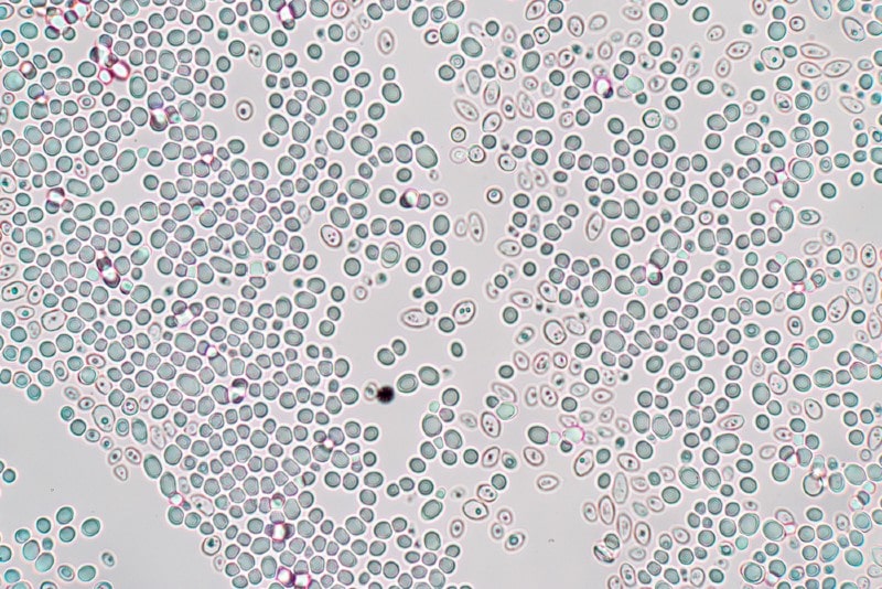
Yeast Cells

Another type of cell you can observe under a microscope is yeast cells. They are single-celled organisms that will appear as egg-shaped organisms under a microscope. In some yeast cells, you might be able to notice the buds as well.
However, to see the organelles inside the yeast cells, you’ll need to practice fluorescence microscopy.
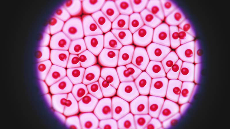
Plant Cells

Most plant cells are somewhat easy to see, which is why it’s good to start with plant cells before observing other cell types. When observing plant cells under a microscope, you should be able to see the nucleus, cytoplasm, cell membrane, and chloroplasts.
For example, for your next experiment, you could look at onion skin to observe the cells inside it. Onion skin has cell walls that our cells don’t have; they are uniform in shape and well-organized. The cell walls you can see on onion skin are constructed from cellulose, protecting the cell and maintaining its shape.
Because of the cell walls, if you don’t stain your specimen, you’ll only be able to see the cell walls under the microscope. However, if you stain the specimen, you will also notice the nucleus.
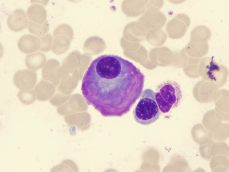
Animal Cells
Animal cells are translucent and colorless, so when you add that to their tiny size, you’ll realize how difficult it’s to see them, even underneath a microscope and especially without staining.
When you observe animal cells, they may appear very different from one another based on the cell type you’re observing. However, their inside structure is pretty much the same. If you compare animal cells to plant cells, you’ll notice that animal cells have a pleomorphic shape because they don’t have cell walls. As there are no cell walls, their shape constantly changes.
The viewing results will be different based on the method you use:
- Direct observing: You should be able to notice the nucleus as a solid structure due to its highest density compared to other cell parts. The membrane should look like a border that closes the inside components of the cell.
- Observing after staining: Observing animal cells after staining will allow you to see much more details than direct observing. You should see the organelles inside the cytoplasm, detailed distinctive parts of the cell, and the nucleus. Under a microscope with high magnification, you should also see ribosomes and mitochondria. In some cells, you should notice chromosomes inside the nucleus, while you can observe structures such as microvilli and cilia when observing tissues.
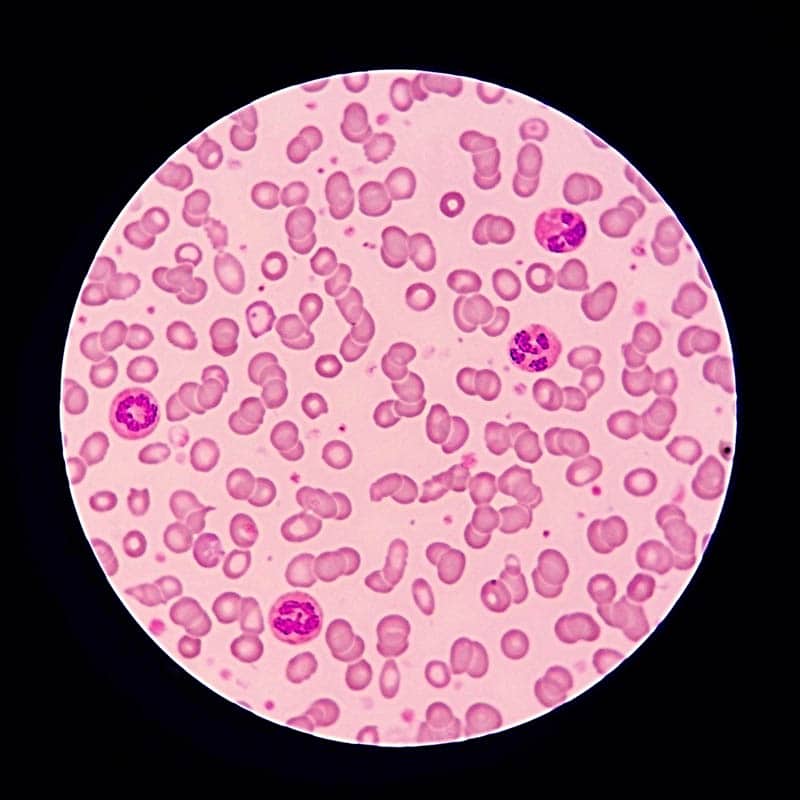
Blood Cells
Our blood contains various components, from white and red blood cells to platelets, etc. However, the highest number of cells inside our bodies are red cells which take over 70% of our cell structure. When observing blood under a microscope, red cells and white cells will stand out the most.
Under a microscope, red cells are small, without a nucleus, and have a doughnut shape. They are thin in the middle, and you can differentiate them from white blood cells, which are significantly larger. They have a nucleus or segmented nuclei; the nucleus divides into smaller parts that are still connected together.

Conclusion
All cells look different under a microscope, so how the cells will look will depend on the type of cell and the way you’re observing them. You can start your experiments with plant cells that are more easily visible, gradually turning to cells that are more difficult to see.
Featured Image Credit: Anamaria Mejia, Shutterstock
About the Author Visnja Radosavljevic
Visnja is a creative, adaptable content writer that covers various topics such as DIY, pets, home improvement, travel, gardening, and more. As a young mom and a college student, she didn’t have enough time to balance her personal and work life, so after multiple years of working a regular 9 to 5 job, she decided to pursue her passion and make a living out of it. She has been writing for a couple of years now, helping people to find valuable and interesting information online.
Related Articles:
How to Clean a Refractor Telescope: Step-by-Step Guide
How to Clean a Telescope Eyepiece: Step-by-Step Guide
How to Clean a Rifle Scope: 8 Expert Tips
Monocular vs Telescope: Differences Explained (With Pictures)
What Is a Monocular Used For? 8 Common Functions
How to Clean a Telescope Mirror: 8 Expert Tips
Brightfield vs Phase Contrast Microscopy: The Differences Explained
SkyCamHD Drone Review: Pros, Cons, FAQ, & Verdict