
Optics Mag is reader-supported. When you buy via links on our site, we may earn an affiliate commission at no cost to you. Read more.
What Does Hair Look Like Under a Microscope? Facts & FAQs
Last Updated on

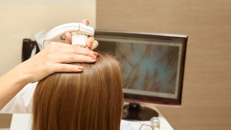
Hair is essentially a protein filament and grows from the follicles that are located in the skin’s dermis. But did you know that all the mammals presently listed on this planet have or have had hair at some point in their lives? And yes, that includes dolphins and whales too. Here’s a second fact that we thought we should share: on average, humans shed about 50 to 150 strands of hair per day.
Hair looks like several things. In some sections, it looks like a tube. While in others, it takes on an oval shape and even creates different patterns. After reading this article, you’ll be able to tell what hair looks like under a microscope, tell apart a healthy strand from a dead one, and the different types of hair that are usually extracted from various animals.

The Different Parts of Hair
Under a microscope, we usually see hair as a structure that is been divided into two parts. There’s the hair shaft and the hair follicle. When you look closely at the shaft, you’ll realize that it’s again divided into three very different layers—the cuticle, cortex, and medulla.
Cuticle
The cuticle is the outermost layer of our hair. It’s made up of several keratin cells that keep on overlapping to protect the internal layer from anything looking to compromise its structural integrity. These cells are naturally transparent, cornified, and ironically, dead. They also happen to have a higher degree of resistance to different chemicals to ensure no outside element finds a way to bind itself to our bodies.
How smooth or shiny your hair looks will hinge on the condition of the cuticles. Maintaining a shiny look is no mean feat, as these cuticles are often subjected to harsh elements all around us. They are always working on overdrive to make sure you’re well protected from excessive heat, ultraviolet light, effects of chlorinated water, aggressive combing, etc.
The funny thing is, we usually assume that conditioning treatments are meant to help us repair our damaged hair. When in reality, they don’t. All they do is add more chemicals onto the layers, thereby making them appear shiny or smooth.
Cortex
The cortex is the prime layer of the hair. We like to view it as the main room where all the extra supplies get stored. For example, if your hair feels the need to hold onto more moisture, this is where it will be stored. If more of its natural pigments need to be hoarded, they’ll also be stored here.
When you examine the cortex layer, you’ll find that it constitutes elongated cells and fusiform (sometimes referred to as spindle-shaped cells). We also noticed it has several air spaces, which are scientifically known as the cortical fusi. Other important features that we can’t ignore are the ovoid bodies and pigment granules.
Your natural hair color will be swayed by the amount of melanin found in the cortex. If, say, your melanin stores are depleted, you’ll have gray hair. And by the way, this is the reason why some hair products on the market are more effective than others. While the sub-standard products target the cuticle, the premium products work extra hard to get to the melanin.

Medulla
Once you get past the cortex, you’ll find the central core, known as the medulla. It’s mostly made up of air bubbles and sparse cells, but we don’t know what role it plays in humans. All we know is that our animals need it in order to regulate their body temperatures. Especially those in regions that experience subzero temperatures.
Human hair doesn’t always come with a medulla. But if you get one that does, and you place it under a microscope, it looks like a black or opaque structure. It could either be a continuous structure or a fragmented one. It all depends on who you got the hair from.

Examining Hair Using a Stereo Microscope

Hair examination is a process just like any other. And that means it’s defined by steps and more importantly, stages. Before you examine the internal structures you must examine the externals using what we call a stereo microscope.
A stereo microscope is also known as a stereo zoom microscope or a dissecting microscope. It’s not as powerful as the compound scope because it has a lower optical resolution power. If you’ve used one before, you know its magnification is never higher than 50X.
Because the stereo microscope has a low resolution it’s often used while examining the general traits of our hair—the texture, shade/color, shape, length, etc.
The procedure is a simple one. Just pick a strand using a pair of tweezers, place it on the stage, configure the scope’s settings, and then keep on adjusting until you get the right focus. The first thing that you’ll take note of is how detailed your hair is. Even with that low-resolution instrument, you’ll still be able to see the hair fibers and fragments.
You’ll also learn that different hairs take on different shapes. Some are straight, some are twisted, some are curly, and so on. The general thickness and the color synthesized by the melanin are all observable traits as well. Go ahead and compare different strands of hair from different sources, to learn all there is to know about this tissue’s physical characteristics.
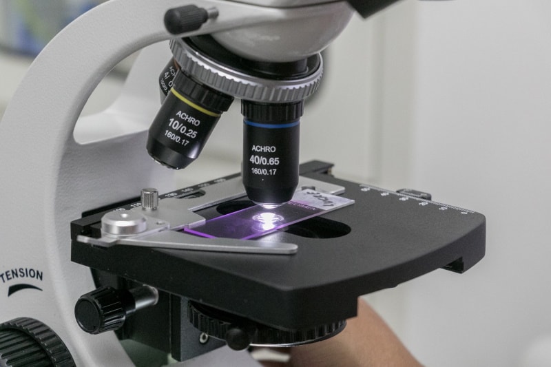
Examining Hair Using a Compound Microscope
To get more insight into your hair’s properties, you’ll need a more powerful instrument, like the compound microscope. And with it, you’ll be able to study both the medulla and the outer layer. Studying the external layer will require the application of a procedure called “scale casting.”
Scale Casting
This procedure entails the utilization of molding material to create a replica of a surface that you’re interested in studying. Specialists in this field like to say that a good scale cast has the ability to preserve the surface texture’s minute details, in a fashion that allows the investigator to easily study it under a microscope.
Preserving the surface texture of your hair’s specimen would be very difficult if this technique never existed. Just think about it; is it possible to effectively examine the cuticle of a specimen when its shape has been distorted?
To be clear, the scale casting procedure is only ideal for the investigation/examination of the hair’s outer layer—the bit that contains the scales. And the microscope’s objectives to be used during this process are the low-power ones.
Biologists like to use this technique to figure out the origins of a specimen. Examining the scale patterns and different shapes usually provides more insight, especially when various types of hairs are compared.
Utilizing a Mount Slide
You’ll have to work with a mount slide if you intend to investigate the internal structure of your specimen. But before you start your examination, ensure the objectives are set to low power. We’re always advised to start with a lower resolution, to find the right focus. It’s still possible to focus with a high-power lens but the margin of error will be wider.
Observing the internal structure of darker hair or one that’s relatively thicker will demand a stronger light intensity.
Frequently Asked Questions (FAQs)
What’s a Medullary Index?
This is what you get once you’ve divided your medulla diameter with the hair’s diameter. The measurements are supposed to be taken using a microscopic ruler, and not your ordinary tools. Animals have a greater medullary index compared to humans, seeing as theirs stands at 0.5. A normal human being will have a medullary index of 0.33.
What’s The Difference Between a Damaged Hair and a Healthy One?
Human hair and animal fur are similar in a lot of ways. For instance, they both look like pigment-filled tubes that are completely covered by several tiny scales. If you want to know whether you’re looking at healthy hair or one that’s damaged, just examine the scales. Those that appear tightly condensed represent healthy hair. Damaged hair has scales that look disheveled.
Why Do Forensic Scientists Analyze Hair?
They analyze hair for several reasons. Let’s say you want to pinpoint the exact origin of a specimen that you just came across. And by “exact” we mean the real owner of the hair. For that to happen, you’ll need to conduct DNA testing on the cells found in the roots. The other type of analysis is one where you perform a chemical study on the sample, to figure out whether the owner has a health condition.

Final Thoughts
Under a microscope, hair looks like several things. In some sections, it looks like a tube. While in others, it takes on an oval shape and even creates different patterns. Providing an accurate description of a strand is virtually impossible since each type of hair is unique to the species. So don’t focus too much on how it looks but try to have fun while studying it!
You Might Also Be Interested In:
Featured Image Credit: Tonhom1009, Shutterstock
About the Author Robert Sparks
Robert’s obsession with all things optical started early in life, when his optician father would bring home prototypes for Robert to play with. Nowadays, Robert is dedicated to helping others find the right optics for their needs. His hobbies include astronomy, astrophysics, and model building. Originally from Newark, NJ, he resides in Santa Fe, New Mexico, where the nighttime skies are filled with glittering stars.
Related Articles:
How to Clean a Refractor Telescope: Step-by-Step Guide
How to Clean a Telescope Eyepiece: Step-by-Step Guide
How to Clean a Rifle Scope: 8 Expert Tips
Monocular vs Telescope: Differences Explained (With Pictures)
What Is a Monocular Used For? 8 Common Functions
How to Clean a Telescope Mirror: 8 Expert Tips
Brightfield vs Phase Contrast Microscopy: The Differences Explained
SkyCamHD Drone Review: Pros, Cons, FAQ, & Verdict