
Optics Mag is reader-supported. When you buy via links on our site, we may earn an affiliate commission at no cost to you. Read more.
What Do Germs & Bacteria Look Like Under a Microscope? Facts & Tips
Last Updated on

Although many people don’t realize this, we come in contact with germs and bacteria every day. We live in a world filled with germs, but it can be hard to understand that if you’ve never seen a germ or bacteria.
That’s where microscopes come in handy, as they allow you to observe germs and bacteria and get a better overview of how they look. Throughout this article, we’ll provide you with more details about germs, bacteria, and what they look like under a microscope.
Click to Skip Ahead:
- What Are Germs and Bacteria?
- What Level of Magnification Do I Need to See Germs and Bacteria?
- What Is a Safe Source of Bacteria You Can Use for Observations?
- Tips on Viewing Germs and Bacteria Under a Microscope
- What Do Germs and Bacteria Look Like?

What Are Germs and Bacteria?
Germs are small microorganisms that can cause health issues and diseases in humans. They commonly spread from one person to the other one through coughing and sneezing, but they can also transmit through body fluids. However, not all are bad, and some bacteria inside us help our bodies absorb nutrients and produce vitamins.
The two most common types of germs are:
- Bacteria: Free-living cells that can survive inside or outside our bodies
- Viruses: Non-living, they require a host to survive
Bacteria represent single-celled organisms that can multiply and live in various environments, even under harsh conditions. They are diverse and can have different structural features.
What Level of Magnification do I Need to See Germs and Bacteria?
Germs and bacteria are tiny, typically around 0.2 micrometers in diameter and around 2 to 8 micrometers in length. That’s why, to see them, you need to observe them under a microscope. However, you need to find the appropriate magnification that will allow you to observe them.
When observing germs and bacteria, it’s easier to observe them in a colony because they only have color in groups. When alone, they are transparent, making it harder to observe them.
Typically, to see the germs and bacteria under a microscope, you need a magnification of 400x to 1000x, but this can vary depending on the type of bacteria and your objective lens. Still, it can be hard to see germs and bacteria even under a microscope.
Why Is It Difficult To See Germs and Bacteria Under a Microscope?
There are several reasons germs and bacteria are difficult to see under the microscope:
- They are tiny: You need a magnification between 400x and 1000x to see germs and bacteria due to their size.
- They are difficult to focus on: Because of the high magnification, the cells can appear out of focus, especially if the water level between the slide and cover glass is thick.
- They are transparent: When bacteria is not in a colony, it will be transparent and therefore barely visible.
- They can be difficult to recognize: Amateurs can have difficulty distinguishing bacteria from dust and dirt on the slide.
What Is a Safe Source of Bacteria You Can Use for Observations?
If you want to observe bacteria for recreational or educational purposes, you need to find a safe source to take your sample. While you should avoid getting bacteria from spoiled food and similar sources such as hummus and soil, you can extract bacteria from safer things, including:
- Cheese: Some cheeses contain bacteria, so they can be an accessible source for a sample you can observe. However, cheese can also have fungi, and those cells can outshine the bacteria.
- Yogurt: It commonly contains cocci bacteria which you should see in chains or pairs.
- Freeze-dried bacteria: You can purchase freeze-dried bacteria and dissolve them in water for observations under a microscope.

The 5 Tips on Viewing Germs and Bacteria Under a Microscope
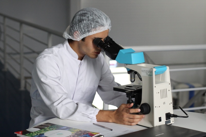
To successfully view germs and bacteria under a microscope, you need to know some tips and tricks to help you with this process. Below is a list of steps you should follow to observe germs and bacteria under a microscope.
1. Prep the Sample
The first thing you need to do is to prepare a sample of the bacteria you want to observe. To do that, you’ll need distilled water and a clean slide. Before dropping water on the slide, ensure your dropper is clean, as the dirt inside could lead to problems when observing the sample.
After placing a couple of drops of distilled water onto the slide, you can add your bacteria. You could also stain the sample by adding safranin or methylene blue, allowing the bacteria to be seen better.
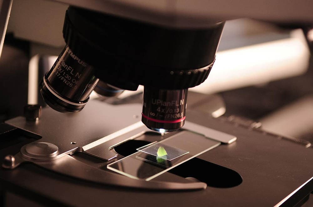
2. Try Different Lens Sizes
To see the bacteria under a microscope, you need to find an appropriate magnification, which can sometimes be challenging. We suggest trying different lens sizes, with at least 400x magnification and up to 1000x magnification.
You need to remember that, although higher magnification will allow you to see more details, it can also make it hard for you to focus the sample. It’s better to start small and increase the magnification level gradually.
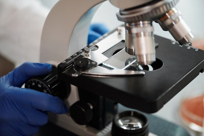
3. Remember Safety

Another thing to remember is that safety comes first, so you should always wear gloves and goggles when handling bacteria. Also, remember to get samples from a safe source and avoid getting samples from spoiled food and body fluids.
4. Look Closely
If this is your first time observing bacteria under a microscope, it might be tricky to see them at first, as they could appear as dirt on the slide. You’ll need to observe the samples intensely to see detailed bacteria.
What Do Germs and Bacteria Look Like?
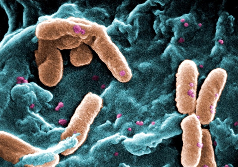
To recognize the bacteria and germs under your microscope, you must know how they look. When it comes to their appearance, bacteria can have three different shapes:
- Cocci (Diplococci, streptococci, tetrad, straphylococci, sarcinae)
- Bacilli (Single bacilus, diplobacilli, streptobacilli, coccobacilli)
- Spiral (Vibrio, spirilla, spirochete)
Cocci Bacteria
These are commonly round-shaped but can be elongated or oval. These bacteria have a unique way of dividing, which is a great way to recognize them under a microscope. Cocci remaining in pairs after dividing are called diplococci, while those that divide and stay connected in a chain are called streptococci.
Bacilli Bacteria
There are fewer bacilli bacteria groups than cocci bacteria, and they are rod-shaped. They divide across their axis, and when they remain in pairs after the division, they are called diplobacilli. However, some bacilli can look like cocci and be oval, which is why they’re called coccobacilli.
In most cases, bacilli will appear as a single cell underneath the microscope, but they can also appear in pairs or chains.
Spiral Bacteria
These bacteria have spirals or twists that set them apart from other bacteria. Vibrios bacteria have a shape that resembles curved rods, while spirilla has a helical shape similar to a corkscrew.
Under a microscope, vibrios will have a comma-like shape while spirilla will be spiral. Spirilla bacteria have flagella on both of their ends, which help them move in aquatic environments. Vibrio bacteria have a flagellum on one end, which allows them to move around.
5. Take Photos of Your Observation
You can photograph your observations when you master the art of finding and distinguishing different bacteria. You can use microscope cameras to capture your findings, which will also help you keep track of all the bacteria you’ve seen and recognized.
Final Thoughts
Although it can be tricky to view germs and bacteria under a microscope, it’s not an impossible task. In fact, if you follow all the tips we provided about how to view them under a microscope, along with how they look, you should be able to notice them quickly. Remember to be patient because your observation skills will get better with time.
Featured Image Credit: Oral bacteria (STEVE GSCHMEISSNER, via Wikimedia Commons CC BY-SA 4.0)
About the Author Visnja Radosavljevic
Visnja is a creative, adaptable content writer that covers various topics such as DIY, pets, home improvement, travel, gardening, and more. As a young mom and a college student, she didn’t have enough time to balance her personal and work life, so after multiple years of working a regular 9 to 5 job, she decided to pursue her passion and make a living out of it. She has been writing for a couple of years now, helping people to find valuable and interesting information online.
Related Articles:
Binocular Magnification Chart: Numbers & Distances Compared
When Were Binoculars Invented? History, Today & Future
How to Clean a Refractor Telescope: Step-by-Step Guide
How to Clean a Telescope Eyepiece: Step-by-Step Guide
How to Clean a Rifle Scope: 8 Expert Tips
Monocular vs Telescope: Differences Explained (With Pictures)
What Is a Monocular Used For? 8 Common Functions
How to Clean a Telescope Mirror: 8 Expert Tips