What Does E. Coli Look Like Under a Microscope?
Last Updated on

It has the power to cause severe diarrhea and vomiting, and is often found in raw, unpasteurized milk, and animal feces. It’s invisible to the naked eye, but what does the mysterious E. Coli look like under a microscope?
Escherichia Coli, named after Theodore Escherich, who discovered the genus in 1885, can appear in food, earth, and the lower intestines of both animals and humans. Some strains of E. Coli cause severe illness, but most of them are harmless.
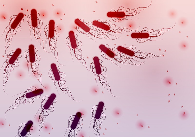
Under a microscope, E. Coli are rod-shaped bacteria that loosely resemble fuzzy capsules. During gram testing, E. Coli bacteria show up as either pink or red in color.

What is Gram Stain Testing?
Gram stain testing is used to distinguish between two large groups of bacteria. Depending on the makeup of the cell walls, the test will result in the bacteria either appearing red or violet.
During the decolorization process of the test, bacterial cells that have a thick peptidoglycan cell wall retain the crystal violet that is used during the staining process. These bacteria are known as gram-positive, and will appear violet. On the other hand, cells with thinner peptidoglycan walls stain red (or pink), as they do not retain crystal violet during the decolorization process.
E.Coli is a gram-negative bacterium, so after gram testing, the capsule-shaped bacterial cells appear either red or pink under the microscope.

How Do You Identify E. Coli Under a Microscope?
Bacteria are usually identified by their characteristics, including their shape and size, and how they behave.
For example, E. Coli is gram-negative, rod-shaped bacilli that measure 2.0–6.0 micrometers in length, and are 1.1–1.5 micrometers in width, with rounded caps on each end. Additionally, they are fast-growing bacteria. Under ideal conditions, E. Coli bacteria can double every 20 minutes.
There are three main types of bacteria:
- Cocci
- Bacilli
- Spiral

- Cocci – These spherical-shaped bacteria are the most common in our environment. They often appear in groups. The term ‘diplococci’ refers to cocci pairs, ‘streptococci’ is when they form a chain, and ‘staphylococci’ refers to clusters of cocci.
- Spiral – As the name suggests, these bacteria are spiral-shaped. Both Campylobacter and Helicobacter are examples of spiral bacteria.
- Bacilli – Bacilli, including E. Coli, are rod-shaped bacteria. As with cocci, bacilli can be found in groups or on their own. Diplobacilli would refer to a pair of bacilli, while streptobacilli would infer chains of bacilli.
Which Type of Microscope is Used to View E. Coli?
Compound light microscopes are great for viewing all kinds of bacteria, skin cells, blood cells, parasites, algae, and tissues. These microscopes are often binocular (they have two eyepieces), and use both optical lenses and light to enhance and enlarge samples.
Compound light microscopes usually have three or four objective lenses that can be rotated into the field of view. These lenses, combined with an eyepiece lens that allows for 10x or 15x magnification, can produce up to a maximum of 1000x magnification.

What Magnification Do You Need to See E. Coli?
You should be able to see E. Coli, and most bacteria, at around 400x magnification. For great detail, 1000x magnification is ideal—anything stronger, however, and it becomes increasingly difficult to keep the moving bacteria in focus.
What Causes E. Coli Infections?
The disease is spread when E. Coli bacteria is ingested, either in contaminated food or drinks. Often, E. Coli infections come about because of unsafe handling of food. Sometimes during the processing, meat can come into contact with bacteria and waste from animal intestines.

What is the Most Likely Source of E. Coli?
According to the U.S. Food and Drug Administration, the primary sources of Shiga toxin-producing E. Coli—a particularly dangerous strain of pathogenic E. Coli—outbreaks are raw or undercooked ground meat products, raw milk and cheeses, and contaminated vegetables and sprouts.
They also advise that E. Coli can also be spread through improper hygiene by food handlers, and even from petting zoos, if the animals are contaminated with pathogenic E. Coli.
The FDA advises everyone to carry out the following measures to protect their households against foodborne illnesses:
Wash Hands
Remember to wash your hands with soap and warm water for at least 20 seconds before and after handling raw foods.

Sanitize Your Kitchen
Wash any cutting boards, countertops, and utensils that may have come in contact with contaminated food. Sanitize them with a bleach and water solution, and dry thoroughly with a clean, unused paper towel.
Clean Your Refrigerator
Wipe up any spills inside your refrigerator immediately. Clean your refrigerator regularly, including the shelves and walls.

Wash Hands After Cleaning
After the cleaning and sanitization process, remember to wash your hands with soap and warm water for at least 20 seconds.
Keep Pet Food Separate
If you have a pet, prevent cross-contamination when preparing their food. As soon as your pets are finished eating, clean up after them and wash their dishes.

Summing Up
Although E. Coli is invisible to the naked eye, pathogenic strains can cause havoc if they enter our digestive systems. The tiny capsule-shaped bacteria can be seen under a microscope at about 400x magnification, where they will appear either as chains or clusters.
At the right temperature, E. Coli can double in numbers in just 20 minutes, making them truly fascinating to study under a microscope.
Featured Image Credit: Piqsels
About the Author Cheryl Regan
Cheryl is a freelance content and copywriter from the United Kingdom. Her interests include hiking and amateur astronomy but focuses her writing on gardening and photography. If she isn't writing she can be found curled up with a coffee and her pet cat.
Related Articles:
How to Clean a Refractor Telescope: Step-by-Step Guide
How to Clean a Telescope Eyepiece: Step-by-Step Guide
How to Clean a Rifle Scope: 8 Expert Tips
Monocular vs Telescope: Differences Explained (With Pictures)
What Is a Monocular Used For? 8 Common Functions
How to Clean a Telescope Mirror: 8 Expert Tips
Brightfield vs Phase Contrast Microscopy: The Differences Explained
SkyCamHD Drone Review: Pros, Cons, FAQ, & Verdict
