What Do the Stages of Mitosis Look Like Under a Microscope? (Images Included)
Last Updated on

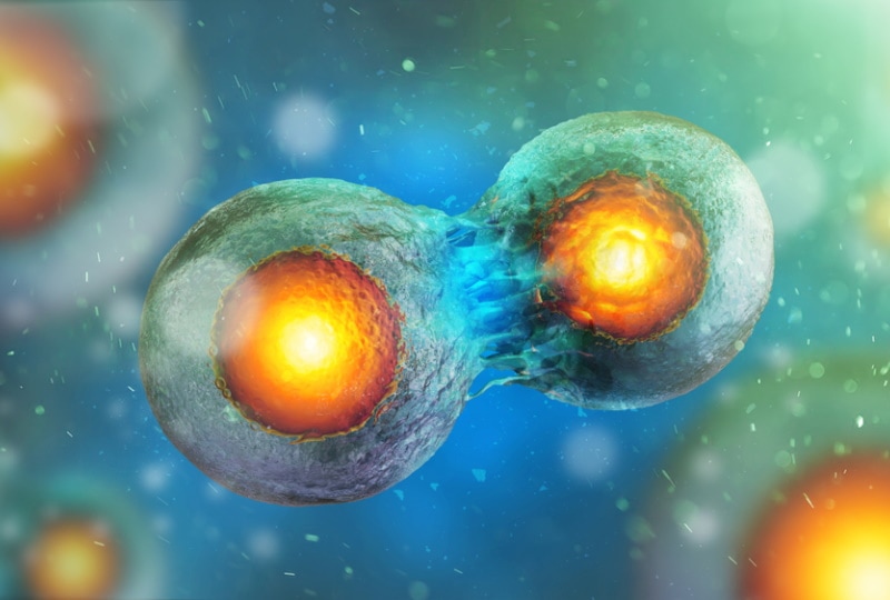
Mitosis is the process of cell division that results in the formation of two daughter cells from a single parent cell. It occurs in all types of cells, including bacteria, archaea, and eukaryotes.
We can see the stages of mitosis under a microscope by staining cells with dyes that bind to different parts of the cell, such as the chromosomes or the cell membrane. But what do these stages look like? Let’s find out.

What Are the Stages of Mitosis?
- Prophase
- Metaphase
- Anaphase
- Telophase
These stages are preceded by interphase, during which the cell grows and replicates its DNA.
Prophase
The chromosomes condense during prophase, becoming visible in the cell. The nucleolus, a structure in the nucleus involved in producing ribosomes, disappears. The nuclear envelope begins to break down. It is a double membrane around the nucleus.
The mitotic spindle also forms during the prophase. It is a structure made up of protein fibers that help separate the chromosomes during mitosis.

Metaphase
During metaphase, the chromosomes line up in the middle of the cell. They are attached to the mitotic spindle by their centromeres, DNA regions connecting the two halves of each chromosome.
Anaphase
In this stage, the mitotic spindle pulls the chromosomes apart, moving them to opposite sides of the cell.
Telophase
The chromosomes go to the opposite poles of the cell and start to de-condense. The nuclear envelope reforms around each set of chromosomes, and the nucleolus reappears.
A new cell wall begins to form between the two daughter cells. The cell then splits in half, creating two new cells.
How Do Stages of Mitosis Appear Under a Microscope?
When viewing mitosis under a microscope, you will see four main stages: prophase, metaphase, anaphase, and telophase. Here’s how they will appear.
Prophase
You will see thick DNA strands coiling and condensing. If you view the cell in the early prophase, the nucleolus may still be intact. It will look like a dark and round blob.
As the prophase progresses, you might see centrosomes on both ends of the cell. Unfortunately, these can be difficult to spot. They look like tiny granules or dots.

Metaphase
You will now be able to make out chromosomes under the microscope. They will form a metaphase plate in the center of the cell. The chromosomes will be attached to spindle fibers at their centromeres. You will also see the mitotic spindle, appearing as if it’s radiating towards the cell’s outer poles.
Anaphase
During anaphase, the chromosomes will move towards the poles of the cell, pulled by the spindle fibers. The process is quick, so you might not see the chromosomes moving if you look at the cell in late anaphase. However, you’ll be able to see the spindle fibers getting shorter as they pull the chromosomes.
Telophase
At telophase, you should be able to see new nucleoli on both ends of the cell. The cell membrane formation is also visible during this step. You’ll see the membrane forming between the daughter cells.

Which Microscopy Techniques Can Be Used to Study Mitosis?

A simple lab microscope won’t do much in studying cell division. But you can use advanced microscopic techniques to observe mitosis.
Brightfield Microscopy
In this type of microscopy, the specimen is illuminated with white light. Therefore, the images produced are usually black and white.
You must stain the cells before observing them under the microscope. Most stains, such as Hoechst 33258 or DAPI, bind to DNA.
Once the cells are stained, you can view them under a microscope and look for signs of mitosis.
Phase-Contrast Microscopy
It is similar to brightfield microscopy but uses a particular lens that makes the cells appear darker or lighter. You can use it to observe living cells since it doesn’t require staining.
Fluorescence Microscope
Fluorescence microscopy uses fluorescent dyes to image living cells. Its principle involves using excitation light to stimulate secondary light emission from the fluorescent dyes.
The emitted light is then captured by a camera and used to generate an image. If you want to study mitosis in living cells, you will use a fluorescent dye that only binds to dividing cells.
The dye will emit light of a specific wavelength that can be detected by the microscope. It lets you follow the stages of mitosis and study how different treatments affect cell division.
Why Do We Need to Study Mitosis?

Mitosis is one of the most critical processes in the life of a cell. It is responsible for the replication and distribution of the cell’s chromosomes and, ultimately, the cell itself.
Without mitosis, cells would be unable to grow or divide, and the body would be unable to repair itself. Therefore, it’s essential to study the stages of mitosis for the following reasons.
Finding Therapeutic Biomarkers
A biomarker is a molecule indicative of a particular disease or condition. The identification of biomarkers is vital in the development of new therapies and treatments for conditions like cancer.
Cancers are caused by the abnormal growth of cells resulting from mutations in the cell’s DNA. These mutations can be caused by various things, including exposure to harmful chemicals or radiation.
By studying the changes that occur in cells during mitosis, scientists may be able to identify biomarkers for cancer. In addition, it would allow the development of new cancer therapies targeting these specific biomarkers.
Treating Birth Defects
Congenital disabilities are often the result of problems during fetal development, including cell division. For example, Down syndrome is caused by the presence of an extra copy of chromosome 21.
The extra chromosome is due to a cell division error called nondisjunction. Scientists can use their knowledge of mitosis to develop treatments for congenital disabilities caused by nondisjunction and other mutations during cell division.

Frequently Asked Questions
Can You See Mitosis With a Microscope?
Yes, you can see mitosis with a microscope. However, you will need a compound light microscope to see mitosis. The high-power objective lens will give you a magnification of 400x, which is enough to see the different stages of mitosis.
What Happens During Mitosis?
In mitosis, a cell divides to form two genetically identical daughter cells. During mitosis, the chromosomes in the nucleus are duplicated, and then the nucleus splits into two. It results in each daughter cell having an identical copy of the chromosomes as the parent cell.
Which Is the Best Microscope to Study Stages of Mitosis?
A fluorescence microscope is the best type of microscope to study stages of mitosis, as it provides the most significant detail. A compound light microscope can also be used, but it will not provide as much detail as a fluorescent microscope.
What Do Chromosomes Look Like Under a Microscope?
Chromosomes appear as thin, thread-like structures under a microscope. They are made up of DNA and proteins and contain a cell’s genetic information.
Are Chromosomes Visible During Mitosis?
You cannot see chromosomes during most of the duration of the cell cycle since they are not condensed. However, they become visible during mitosis due to condensation. You can see them as distinct dark bodies.
What Is Another Name for Mitosis?
Mitosis is also called equatorial division because the chromosomes line up in the middle of the cell during this stage.

Final Thoughts
Mitosis is an important biological process as it helps ensure the survival of a species by allowing cells to divide and replenish themselves. The four stages of mitosis include prophase, metaphase, anaphase, and telophase.
When observing mitosis under a microscope, you can see the different stages of cell division happening. The chromosomes appear as long, thin strands during prophase.
During metaphase, the chromosomes form a metaphase plate in the center of the cell, while in anaphase, they move to the opposite ends. While you can use brightfield microscopy to study mitosis, fluorescent microscopy gives more detail and can be used to label specific chromosomes or proteins of interest.
Featured Image Credit: Andrii Vodolazhskyi, Shutterstock
About the Author Jeff Weishaupt
Jeff is a tech professional by day, writer, and amateur photographer by night. He's had the privilege of leading software teams for startups to the Fortune 100 over the past two decades. He currently works in the data privacy space. Jeff's amateur photography interests started in 2008 when he got his first DSLR camera, the Canon Rebel. Since then, he's taken tens of thousands of photos. His favorite handheld camera these days is his Google Pixel 6 XL. He loves taking photos of nature and his kids. In 2016, he bought his first drone, the Mavic Pro. Taking photos from the air is an amazing perspective, and he loves to take his drone while traveling.
Related Articles:
How to Clean a Refractor Telescope: Step-by-Step Guide
How to Clean a Telescope Eyepiece: Step-by-Step Guide
How to Clean a Rifle Scope: 8 Expert Tips
Monocular vs Telescope: Differences Explained (With Pictures)
What Is a Monocular Used For? 8 Common Functions
How to Clean a Telescope Mirror: 8 Expert Tips
Brightfield vs Phase Contrast Microscopy: The Differences Explained
SkyCamHD Drone Review: Pros, Cons, FAQ, & Verdict
