What Microscope Has the Highest Magnification? The Answer is Fascinating!
Last Updated on

Microscopy has come a long way since the invention of the first microscope decades ago. Today, microscopes allow us to magnify a specimen up to 50 million times.
As a result, microscopes have become an invaluable tool in science, providing researchers with a way to examine specimens in unprecedented detail. So, which microscope has the highest magnification? Electron microscopes are the most powerful microscopes available today and can magnify a specimen up to 50 million times.
Of course, the level of magnification you need will depend on the application you have in mind. However, for most purposes, a lower magnification microscope will suffice. Here’s a closer look at the working of electron microscopes and what they can allow you to see.

Which Type of Microscope Is the Most Powerful?
In terms of microscopy, power refers to the magnification capability of a microscope. The higher the power, the greater the magnification. There are several microscopes, each with its inherent level of power. However, electron microscopes have the highest magnification.
They accomplish this by using a beam of electrons instead of light to create an image. It gives them a much higher resolution than other microscopes and allows them to show finer details.

Which Is the Highest Magnification Image Ever Taken?
The highest magnification image ever was taken in 2009 by scientists in Zurich. They used non-contact atomic force microscopy to take a picture of a pentacene molecule. The microscope has a cantilever with a very sharp tip that is used to scan the surface of an object. As a result, the cantilever tip’s response is used to generate the image.
Pentacene is a molecule made up of five fused benzene rings. The image taken by the microscope showed individual atoms on the pentacene molecule. The hydrocarbon has a length of 1.4 nanometers, 500,000 times smaller than a single hair on your head.
How Do Microscopes Magnify Objects?
The magnification power of a microscope is determined by its optical system, which consists of several lenses that focus light onto the specimen. The power of the objective lens, which is the lens closest to the specimen, is the most crucial factor in determining the overall magnification power of the microscope. If the objective lens has low power, such as 10x, the microscope will have a low magnification power. On the other hand, an objective lens with high power, such as 100x, will result in a high magnification power.
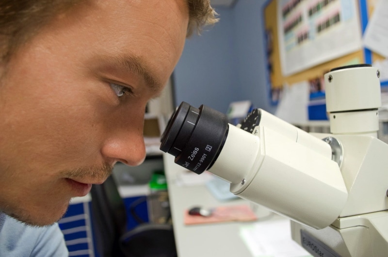
In a simple light microscope, light enters the objective lens from below and is focused onto the specimen. It then passes through the specimen and is focused again by the objective lens onto the ocular lens, which magnifies the image and projects it onto the eye’s retina.

The total magnification power of the microscope is equal to the power of the objective lens multiplied by the power of the ocular lens. For example, if the objective lens has a power of 10x and the ocular lens has a magnification of 4x, the total magnification power of the microscope would be 40x.
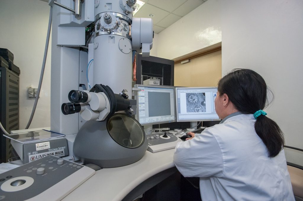
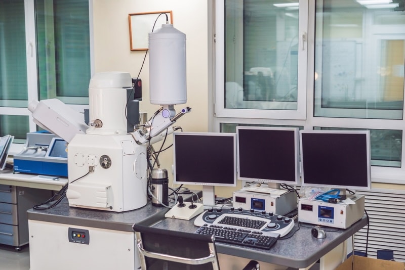
Meanwhile, electron beams are used instead of light in an electron microscope to produce a magnified image of the specimen. The electron beams are generated by an electron gun and are focused by an electromagnetic lens onto the specimen. The beams then pass through the specimen and are focused again by the lens onto an electronic detector. Finally, it converts the beams into an electrical signal. The electrical signal is amplified and displayed on a screen, producing a magnified specimen image.
What Is the Smallest Thing You Can See With a Microscope?
The smallest thing you can see with a microscope is an atom. An atom’s size is nearly 0.1 nanometer. You can use a scanning tunneling microscope, a type of electron microscope, to view and photograph atoms. Meanwhile, the smallest size you can see is 500 nanometers if you’re using a high-quality light microscope. To put things in perspective, one nanometer is one billionth of a meter. Thus, 500 nanometers means the specimen is 200 times smaller than a human hair’s width. For example, bacteria may range up to 1,000 nanometers in size.


Final Thoughts
Electron microscopes have the highest magnification. In addition, the transmission electron microscope has the highest resolving power of the two types of electron microscopes, while the scanning electron microscope produces better three-dimensional images.
The two types of microscopes also have different applications. For example, the transmission electron microscope is used to study the structure of materials at the atomic level. In contrast, the scanning electron microscope helps researchers learn about the surface features of the specimen.
The smallest things we can see with electron microscopes are proteins, cellular structures, and even individual atoms.
Featured Image Credit By: Piqsels
About the Author Jeff Weishaupt
Jeff is a tech professional by day, writer, and amateur photographer by night. He's had the privilege of leading software teams for startups to the Fortune 100 over the past two decades. He currently works in the data privacy space. Jeff's amateur photography interests started in 2008 when he got his first DSLR camera, the Canon Rebel. Since then, he's taken tens of thousands of photos. His favorite handheld camera these days is his Google Pixel 6 XL. He loves taking photos of nature and his kids. In 2016, he bought his first drone, the Mavic Pro. Taking photos from the air is an amazing perspective, and he loves to take his drone while traveling.
Related Articles:
What Is the Best Binocular Magnification for Hunting? Optical Features Explained
How to Clean a Refractor Telescope: Step-by-Step Guide
How to Clean a Telescope Eyepiece: Step-by-Step Guide
How to Clean a Rifle Scope: 8 Expert Tips
Monocular vs Telescope: Differences Explained (With Pictures)
What Is a Monocular Used For? 8 Common Functions
How to Clean a Telescope Mirror: 8 Expert Tips
Brightfield vs Phase Contrast Microscopy: The Differences Explained
