
Optics Mag is reader-supported. When you buy via links on our site, we may earn an affiliate commission at no cost to you. Read more.
Can Viruses Be Seen With a Light Microscope? The Interesting Answer!
Last Updated on

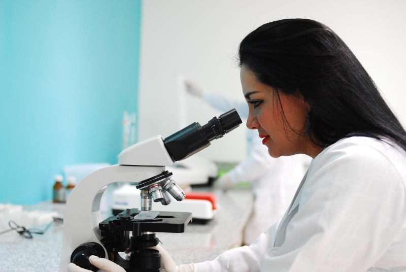
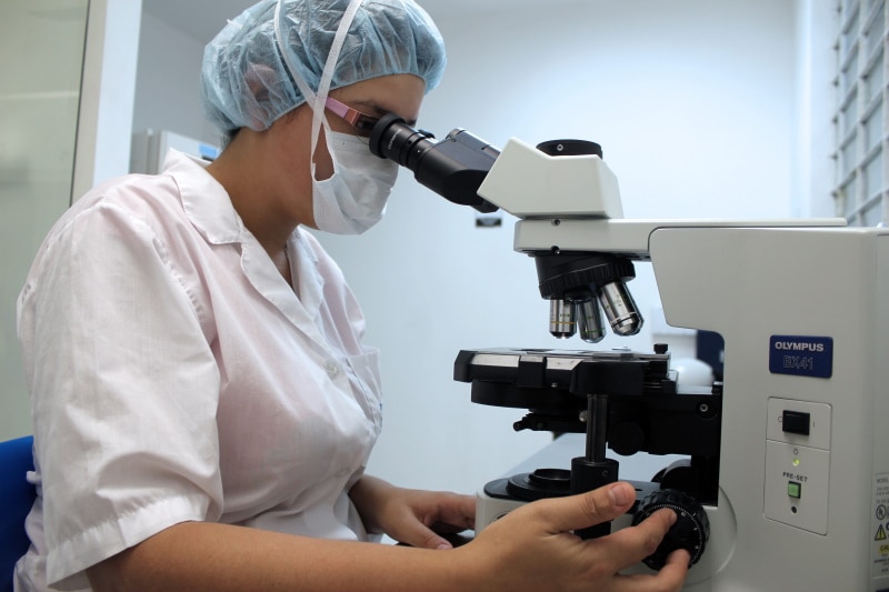
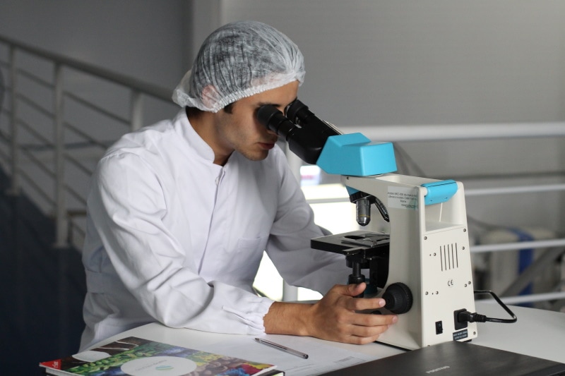
A light microscope is a powerful tool for analyzing fungi or parasites. However, it cannot be used to study viruses because viruses are much smaller than these other organisms.
A virus can be as small as 0.02–0.3 micrometers (μm), which is about one-hundredth to one-thousandth the size of a bacterium. To put this into perspective, a micrometer is one-millionth of a meter or about one-twenty-fifth of the diameter of a human hair.
Because they are so tiny, you cannot see viruses with a light microscope. Instead, you have to use an electron microscope. Here’s a deeper dive into the study of viruses.

Which Microscopes Are Used to View Viruses?
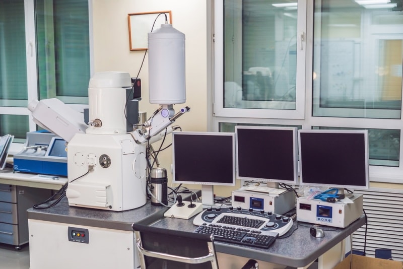
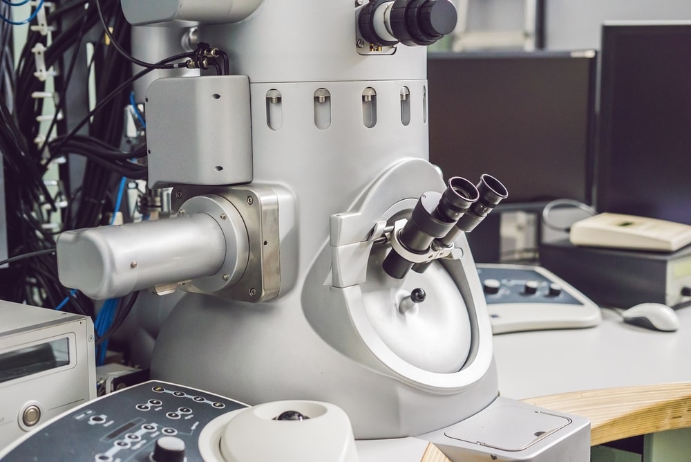
Electron microscopes can obtain very high-resolution images of viruses and other similar particles. An electron microscope uses a beam of electrons to produce an image of the specimen rather than light waves.
Since electrons have a much shorter wavelength than light waves, this allows for a much higher resolution. However, viruses are typically only a few nanometers, so even the best optical microscopes can only provide limited information about them.
Two types of electron microscopes are commonly used to study viruses: the transmission electron microscope (TEM) and the scanning electron microscope (SEM).
Transmission Electron Microscope
A transmission electron microscope (TEM) is an electron microscope that uses a beam of electrons to create an image of the specimen. The specimen is often an ultrathin section less than 100 nanometers thick.
To be able to visualize the structure of the specimen, it must be electrically conductive. The image is formed by the interaction of the electrons with the sample as they pass through it.
The resolving power of the TEM is typically much greater than that of the light microscope, allowing it to image details as small as 0.1 nanometers in diameter.
Scanning Electron Microscope
An SEM differs from a TEM because it does not use a beam of electrons to create an image. Instead, it uses a focused beam of electrons to scan the surface of the specimen.
As the beam moves across the specimen, it produces a signal converted into an image. The SEM can provide information about the specimen’s topography and elemental composition. Because the SEM does not require a conductive specimen, researchers can use it to study a wide variety of materials, including biological samples.
Who Studies Viruses?
Typically, a virologist studies viruses to understand how they interact with their host. For example, if we can understand how a virus makes people sick, we may be able to develop treatments or vaccines.
Besides researching how viruses cause disease, virologists also work to understand how viruses evolve. Depending on their specialization, virologists may work in a laboratory or field. For example, fieldwork for a virologist could involve collecting samples from sick people or animals.
Scientific researchers and pathologists might also study viruses. The research around viruses involves studying their genetic material, structure, and lifecycle. In addition, experimentation is important to learn more about how a virus interacts with cells.
Techniques Used to View Viruses Under a Microscope
When using electron microscopes to image viruses, there are generally two main techniques: negative staining and electron tomography.
Negative staining involves using a stain that will not interact with the virus but will bind to the background material. As a result, the virus appears as a clear or white object against a darker background.
Electron tomography is a three-dimensional imaging technique that can show viruses within their native environment, such as within a cell. It involves taking a series of two-dimensional images at different angles and piecing them together to create a three-dimensional image.
Besides these staining techniques, researchers also use different microscope methods to view viruses.
Fluorescence Microscopy
In this method, viruses are first tagged or labeled with fluorescent molecules. These can be either small organic molecules or antibodies binding to the virus.
The viruses are then exposed to a specific wavelength that causes the fluorescent molecules to emit light of a different wavelength. The emitted light is then detected and captured by a camera, resulting in an image of the viruses.
Use: Fluorescence microscopy helps in quantitative estimation. For example, if you want to know how many virus particles infect a particular cell, you can count them using this method.
Total Internal Reflection Dark-Field Microscopy (TIRDFM)
Total Internal Reflection Dark-Field Microscopy is a newer technique that uses light to image viruses. In this method, a specialized microscope lens is used to illuminate the sample from below. It creates an evanescent field, a light field that extends just a few tens of nanometers from the sample’s surface. You do not need to label the virus with fluorescent molecules for this method. The viruses reflect the light, and the microscope captures the reflection to create an image.
Use: TIRDFM can be used to image viruses that are difficult to label with fluorescent molecules, such as those that do not have a surface protein that an antibody can target.
Transmission Electron Microscopy
Offering a 1,000 times higher resolution than light microscopy, Transmission Electron Microscopy (TEM) is one of the most widely used techniques for imaging viruses. In this method, a beam of electrons passes through a thin sample, such as a virus suspended in water. The electrons interact with the atoms in the sample, and this interaction creates an image.
TEM can be used to image viruses at different stages of their life cycle, such as when they are inside or outside of a cell.
Use: TEM can be used to study the structure of viruses in great detail. It is also used to study how viruses interact with cells and determine viruses’ three-dimensional structure.
Cryo-Electron Microscopy
Any technique that has something to do with “cryo” involves very low temperatures, and Cryo-Electron Microscopy (Cryo-EM) is no different.
In Cryo-EM, viruses are first frozen, so they are in a state of arrested development. They are then placed on a metal grid and bombarded with electrons. The electrons interact with the atoms in the sample, helping the microscope create an image. The researcher may reconstruct the images to form a three-dimensional viral structure.
Use: Cryo-EM is a helpful technique for studying the attachment of viruses to cells. For example, researchers have used it to study how the Zika virus attaches to cells in the placenta. Likewise, you can use it to study viral replication and identify different mechanisms during the process.
Why Do We Need to Study Viruses Under a Microscope?
Virology is the study of viruses and the diseases they cause. It is a relatively new science, only emerging as its field in the early twentieth century.
The Covid-19 pandemic should be enough to illustrate the importance of studying viruses. However, there are many other reasons why viruses are essential to our understanding of the world and how it works. Some reasons to study viruses include:

- Study Diseases: Viral pathologies are some of the most difficult to treat. We need a thorough understanding of how viruses work and what makes them tick to develop treatments and vaccines.
- Understand Evolution: Viruses are some of the simplest life forms on Earth and can help us understand how more complex organisms came to be.
- Learn About Immunity: Our bodies have evolved ways to fight viral infections. By understanding how viruses work, we can learn more about how our immune system works and how to strengthen it.
- Protect Against Bioterrorism: Viruses can be weaponized and used as bioweapons. Therefore, it is important to study them so we can be prepared for a bioterrorist attack.
While the Covid-19 pandemic has brought viruses to the forefront, they have been studied for centuries. The invention of the electron microscope allowed scientists to take a closer look at viruses and understand how they work.
For instance, we can now purify viruses from their organelles and cellular proteins. Then, we can study the basic steps of viral replication.

Final Thoughts
A light microscope cannot be used to view or study viruses. Instead, you need an electron microscope to see viruses. Electron microscopes use a beam of electrons to create an image of the specimen.
The inability of the light microscope to show viruses is due to the small sizes of the organisms and the low magnification power of the microscopes. However, we can use different electron microscopy techniques, such as Cryo-EM, to study the viral structure and function.
Studying viruses is important to understand their role in diseases and how we can develop treatments and vaccines for viral infections.
Featured Image Credit: Artem Podrez, Pexels
About the Author Jeff Weishaupt
Jeff is a tech professional by day, writer, and amateur photographer by night. He's had the privilege of leading software teams for startups to the Fortune 100 over the past two decades. He currently works in the data privacy space. Jeff's amateur photography interests started in 2008 when he got his first DSLR camera, the Canon Rebel. Since then, he's taken tens of thousands of photos. His favorite handheld camera these days is his Google Pixel 6 XL. He loves taking photos of nature and his kids. In 2016, he bought his first drone, the Mavic Pro. Taking photos from the air is an amazing perspective, and he loves to take his drone while traveling.
Related Articles:
What Is the Best Binocular Magnification for Hunting? Optical Features Explained
Can You Use Binoculars to Look At Stars? How to Choose the Right Pair
How to Clean a Refractor Telescope: Step-by-Step Guide
How to Clean a Telescope Eyepiece: Step-by-Step Guide
How to Clean a Rifle Scope: 8 Expert Tips
Monocular vs Telescope: Differences Explained (With Pictures)
What Is a Monocular Used For? 8 Common Functions
How to Clean a Telescope Mirror: 8 Expert Tips