
Optics Mag is reader-supported. When you buy via links on our site, we may earn an affiliate commission at no cost to you. Read more.
Which Microscope Is Best for Metal Surfaces? What to Know!
Last Updated on

A scanning tunneling microscope is used to create high-resolution images of the surfaces of non-conducting materials. Therefore, it is best suited for imaging metal surfaces. Besides choosing a suitable microscope, selecting the proper technique for viewing the specimen is also essential.
Some techniques used to study metal surfaces include x-ray photoelectron spectroscopy, atomic force microscopy, and electron microscopy. In this article, we go into detail about viewing metal surfaces under a microscope and the kind of microscope best suited for the job.

Which Microscope Can You Use to Study Metal Surfaces Under a Microscope?
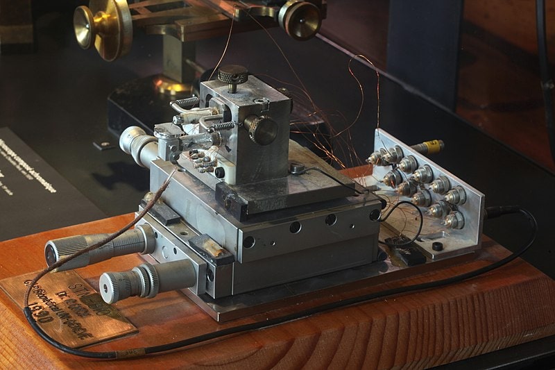
A scanning tunneling microscope is the best type of microscope to use to study metal surfaces. The microscope uses a needle to scan the surface of the metal and create an image.
The needle is attached to a cantilever mounted on a piezoelectric crystal. The crystal vibrates the cantilever, and the needle is moved across the material’s surface.
The image is then magnified so you can see the details of the surface. The resolution of this microscope is very high. Thus, you can see even the most minor details of the metal surface.
Top 4 Techniques Used to View Metals Under a Microscope
Nowadays, scientists use various techniques to study metal surfaces under a microscope. However, each method has its advantages and disadvantages.
Some of the most popular techniques are as follows.
1. X-Ray Photoelectron Spectroscopy
X-ray photoelectron spectroscopy (XPS) is a technique that uses X-rays to study the surface of a metal. In this technique, x-rays bombard a metal surface. These X-rays cause the electrons in the metal to be excited. As these electrons return to their original energy levels, they emit photons.
An XPS machine can detect these photons and create a map of the metal’s surface. X-ray photoelectron spectroscopy is very sensitive to the chemical composition of the surface, but it can only be used on thin samples.
Why Use It?
- XPS is a sensitive technique and can detect even minimal changes in the surface of a metal.
- It can also be used to study the composition of a metal’s surface.
2. Scanning Electron Microscopy
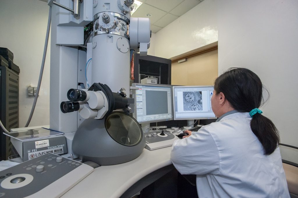
A scanning electron microscope (SEM) uses electrons to create an image of the surface of a metal. When a metal is in an SEM, it’s bombarded with a beam of electrons.
These electrons interact with the atoms on the surface of the metal, causing them to emit photons. Essentially, photons are a form of light, so by detecting these photons, an image of the metal’s surface can be created.
Why Use It?
- SEM is a very versatile technique used to study a wide range of metal surfaces.
- It is also relatively easy to use, which makes it a popular choice for scientists.
3. Atomic Force Microscopy
Atomic force microscopy (AFM) is a popular technique for studying metal surfaces. The method uses a sharp tip to scan the surface of the metal.
The force between the tip and the surface is used to generate a topographic image of the metal. Here is how the process goes:
- First, you need to place the metal sample on a particular stage. Then, you position the microscope’s tip above the specimen.
- Once the tip is in position, you apply a small amount of force to the tip. Doing this will cause the tip to come into contact with the sample’s surface.
- The force between the tip and the surface will be measured as the tip moves across the surface. The microscope will then use this information to generate a topographic image of the surface.
A topographic image is a three-dimensional image showing the surface’s height.
Why Use It?
- The main advantage of AFM is that you can use it to study a wide range of metal surfaces.
- In addition, AFM can be used to study surfaces at the atomic level.
4. Light Microscopy
Another technique researchers commonly use to study metals in light microscopy. Light microscopes use visible light and magnifying lenses to enlarge images of small objects. The method helps explore the surface features of metals.
Here are the standard light microscopy methods for metallurgical studies:
- Brightfield Microscopy: In this method, white light illuminates the dark specimen. The image appears dark against a bright background.
- Darkfield Microscopy: In darkfield microscopy, the specimen is illuminated with light blocked by a circular stop or diaphragm. As a result, you see a bright image against a dark background.
- Crossed Polarizers: Polarizers are placed at right angles to each other in front of the eyepiece and light source. The method helps study birefringent specimens, such as metals. Birefringent materials have different indices of refraction for light traveling in different directions.
- Nomarski Differential Interference Contrast: NDIC is similar to crossed polarizers, but a compensator is present in place of one of the polarizers. The compensator creates areas of interference that appear as a contrast in the image.

Why Use It?
- Light microscopy has various applications in different industries.
- You can use multiple light microscopy methods based on your particular application.
Mechanism of Viewing Metals Under a Microscope
Before understanding how it’s possible to view metals under a microscope, it’s essential to understand the structure and composition of metals.
Metals have a crystalline structure, meaning the atoms are arranged in a regular pattern. The pattern is known as a lattice. The atoms in a metal are held together by strong electrical forces. The electrons create these forces in the atoms. The electrons in the atom’s outermost energy level are not held as tightly to the nucleus as the electrons in the inner orbits.
The electrons in the outer level can move freely from one atom to another. This is what gives metals their electrical conductivity. The electrons in the outer energy level are also responsible for the metallic luster. It refers to the shiny appearance that metals have. So how does this structure allow metals to be viewed under a microscope?
The answer has to do with the way that light interacts with metals. When light shines on a metal, the free electrons in the outer energy level absorb some of the energy from the light. They then re-emit that energy as light. The process is known as fluorescence. Thus, a microscope with a particular light source can help view metals. The light source shines a light on the metal, and the free electrons absorb the energy and re-emit it as light. The light can then be captured by the microscope and magnified to allow us to see the metal on a much smaller scale.

Applications of Viewing Metals Under a Microscope
The ability to view metals under a microscope has many applications. Here are some of them.
Metallography
Metallography is the study of the microstructure of metals. The microstructure is the organization of the metal at the atomic level. For instance, it shows the arrangement of atoms, how they interact with each other, and what kind of defects are present.
We need this information to understand how different metals behave under different conditions. For example, when we heat metal, the atoms vibrate more, and this can cause the metal to change shape. We can use metallography to study how different metals will respond to this change in temperature.
Subsequently, the information will find use in different industries, such as aerospace, automotive, and energy.
Forensics
Forensic scientists may use a microscope to examine metal evidence from a crime scene. It could include anything from a murder weapon to a piece of jewelry.
The scientist can use the microscope to look for unique markings that identify the item’s manufacturer. Likewise, researchers can use it to match the metal evidence to a specific event.
In this way, they can connect a suspect to a crime.

Laser System
Many different types of lasers are used in industrial and medical settings. But how do we know if the laser is working correctly?
By looking at the metal under a microscope, we can see how the laser is affecting the metal. The information is essential for developing new lasers and improving existing ones.
Conclusion
You can use a scanning tunneling microscope to study metals under a microscope. Some techniques for studying metals include light, atomic force, and electron microscopy.
All three of these techniques have their merits and demerits, so it is vital to choose the right one, depending on your needs.
Speaking of needs, the microscopic study of metals has many applications in forensics, engineering, and medicine. With the development of new techniques, we can expect to see even more applications in the future.
Featured Image Credit: Piqsels
About the Author Jeff Weishaupt
Jeff is a tech professional by day, writer, and amateur photographer by night. He's had the privilege of leading software teams for startups to the Fortune 100 over the past two decades. He currently works in the data privacy space. Jeff's amateur photography interests started in 2008 when he got his first DSLR camera, the Canon Rebel. Since then, he's taken tens of thousands of photos. His favorite handheld camera these days is his Google Pixel 6 XL. He loves taking photos of nature and his kids. In 2016, he bought his first drone, the Mavic Pro. Taking photos from the air is an amazing perspective, and he loves to take his drone while traveling.
Related Articles:
What Is the Best Binocular Magnification for Hunting? Optical Features Explained
How to Clean a Refractor Telescope: Step-by-Step Guide
How to Clean a Telescope Eyepiece: Step-by-Step Guide
How to Clean a Rifle Scope: 8 Expert Tips
Monocular vs Telescope: Differences Explained (With Pictures)
What Is a Monocular Used For? 8 Common Functions
How to Clean a Telescope Mirror: 8 Expert Tips
Brightfield vs Phase Contrast Microscopy: The Differences Explained